
Fig. 1
- ID
- ZDB-IMAGE-060727-11
- Publication
- Hammerschmidt et al., 1996 - dino and mercedes, two genes regulating dorsal development in the zebrafish embryo
- All Figures
- Figures for Hammerschmidt et al., 1996
|
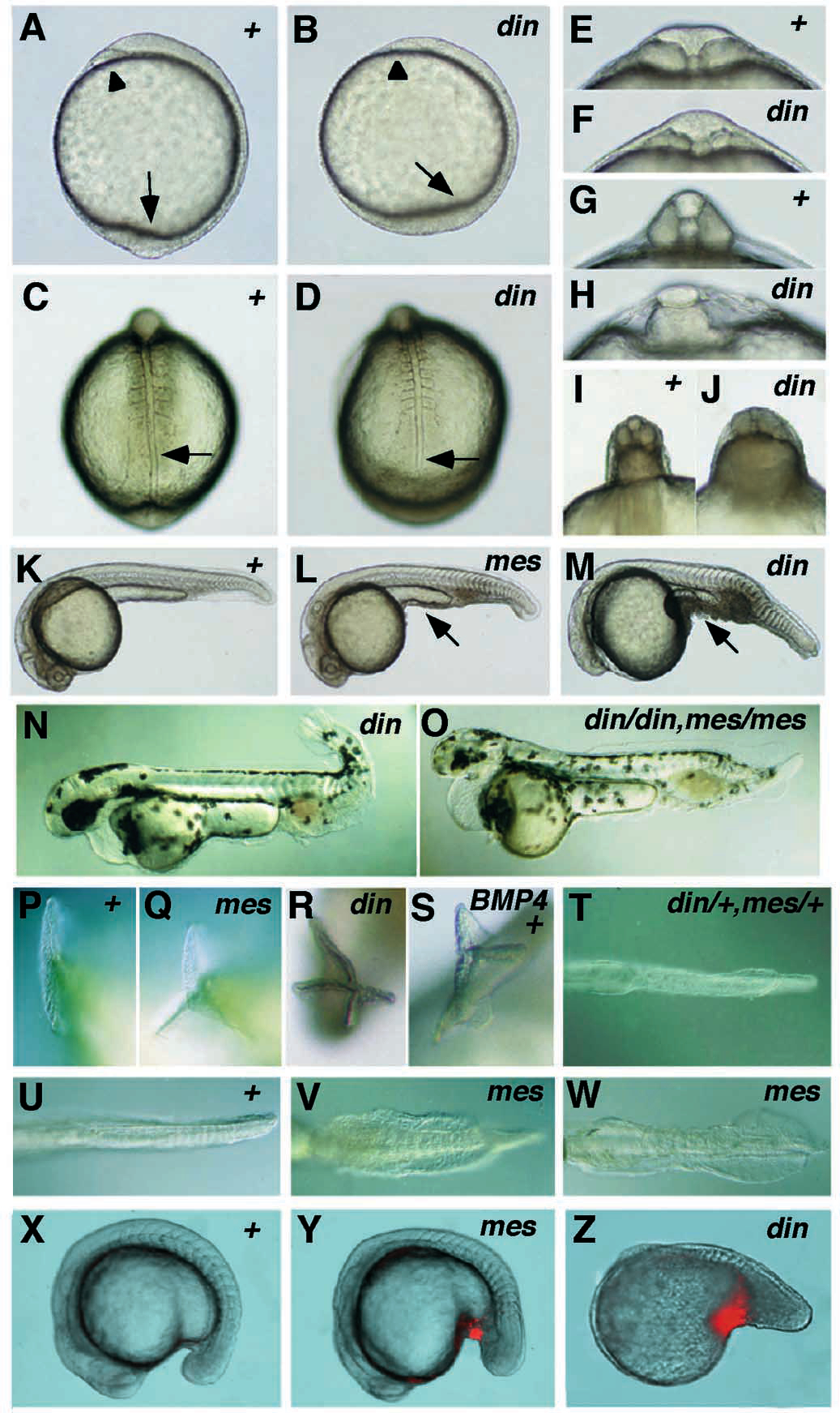
Fig. 1 Morphological characteristics of the dino (dintm84) and mercedes (mestm305) mutant embryos. Wild-type embryos are indicated with ‘+’. (A,B) Tailbud stage, lateral view: the pillow is marked with an arrowhead, the tailbud with an arrow. (C,D) 8-somite stage, dorsal view: the end of the notochord and the corresponding position in the wild-type embryo are marked by an arrow. (E,F) 3- somite stage, optical cross section at the level of the 2nd somite: the anterior somites of the mutant are smaller and the neural tube is of normal size. (G,H) 8-somite stage, optical cross section at the level of the 6th somite: the notochord is lost and the somites are fused in dino mutants with a strong phenotype. (I,J) 15-somite stage, optical cross section at the level of the 13th somite: the posterior somites are larger and the neural tube is smaller in dino mutants. (K,L,M) 24 hours, lateral view: cells at the ventral side of the yolk extension are marked with an arrow. (N,O) 48 hours, lateral view: note the smaller head in the din mes double mutant. (P-S) 36 hours, posterior view of tail fin; (S) wild-type embryo injected with Xenopus Bmp-4 mRNA. (T-W) 36 hours, ventral view of tail fin: (V) mestm305/mestm305 embryo from heterozygous mother; (W) mestm305/mestm305 embryo from homozygous mother. (X-Z) Lateral views of (X) wild-type, 20- somite stage embryo; (Y) mestm305, 20-somite stage embryo; (Z) dintt250, 15-somite stage embryo, showing acridine orange staining. Apoptotic cells (red) can be detected ventral of the forming yolk extension in din and mes mutant embryos.