
|
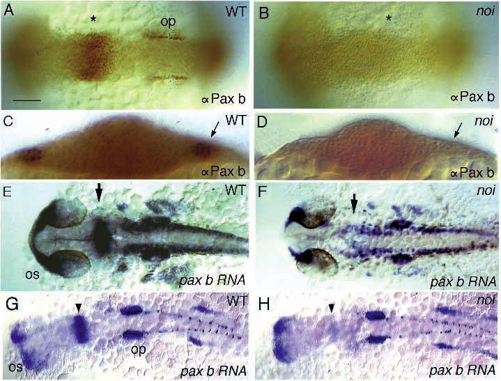
Fig. 5 Pax-b protein, but not RNA expression, is affected in noi. Shown are dorsal views of wild-type and noi mutant embryos (axial view as optical section in C and D), stained for pax-b protein and RNA, as indicated. (A-D) pax-b antibody staining is eliminated from (A,B) the MHB primordium (asterisks) and otic placode (op) and (C,D) the pronephric anlage (arrow) of mutant embryos at the 5-somite stage. (E,F) pax-b RNA expression in wild type and noith44 mutant embryos. RNA expression is found in all tissues of the mutant, except for the MHB (arrow) and pronephros. Notice the overall reduction of RNA level in the mutant, which is not yet apparent at the 14-somite stage in (G,H): only a trace of RNA can be detected in the MHB of mutant embryos at this stage (arrowhead), whereas RNA expression in the optic stalk (os), otic placode, pronephric anlage and hindbrain neurons is normal. Scale bar, 100 µm (A,B); 50 µm (C,D); 140 µm (E-H).