
|
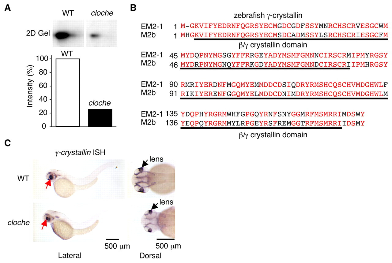
Fig. 1 Crystallin proteins levels are diminished in cloche embryos. (A) 2D-gel electrophoresis: Total 2.5 dpf extracts as described in the Materials and methods were analyzed. A spot (20 kDa, pI of 8.8) was identified that was strongly reduced in cloche compared with wild-type (WT) embryos. Spot intensities were analyzed by an image analysis program (ImageJ) and the values were normalized to wild type (100%). (B) The spot was cut out of the gel and identified by MS/MS spectrometry sequencing to be a member of the γ-crystallin family. This protein, designated as embryonic γ-crystallin M2 type1 (EM2-1) was 78% homologous to a recently described zebrafish γ-crystallin M2b (M2b) that had been cloned from adult lens tissue (Wistow et al., 2005). The two sequences were aligned. Identical amino acids are in red. The Reverse Position Specific BLAST program showed two ß/γ-crystallin domains that are underlined in black. (C) γ-crystallin gene expression was analyzed by in situ hybridization at 2.5 dpf. Both lateral and dorsal views are shown. γ-crystallin is expressed solely in the lens.