
|
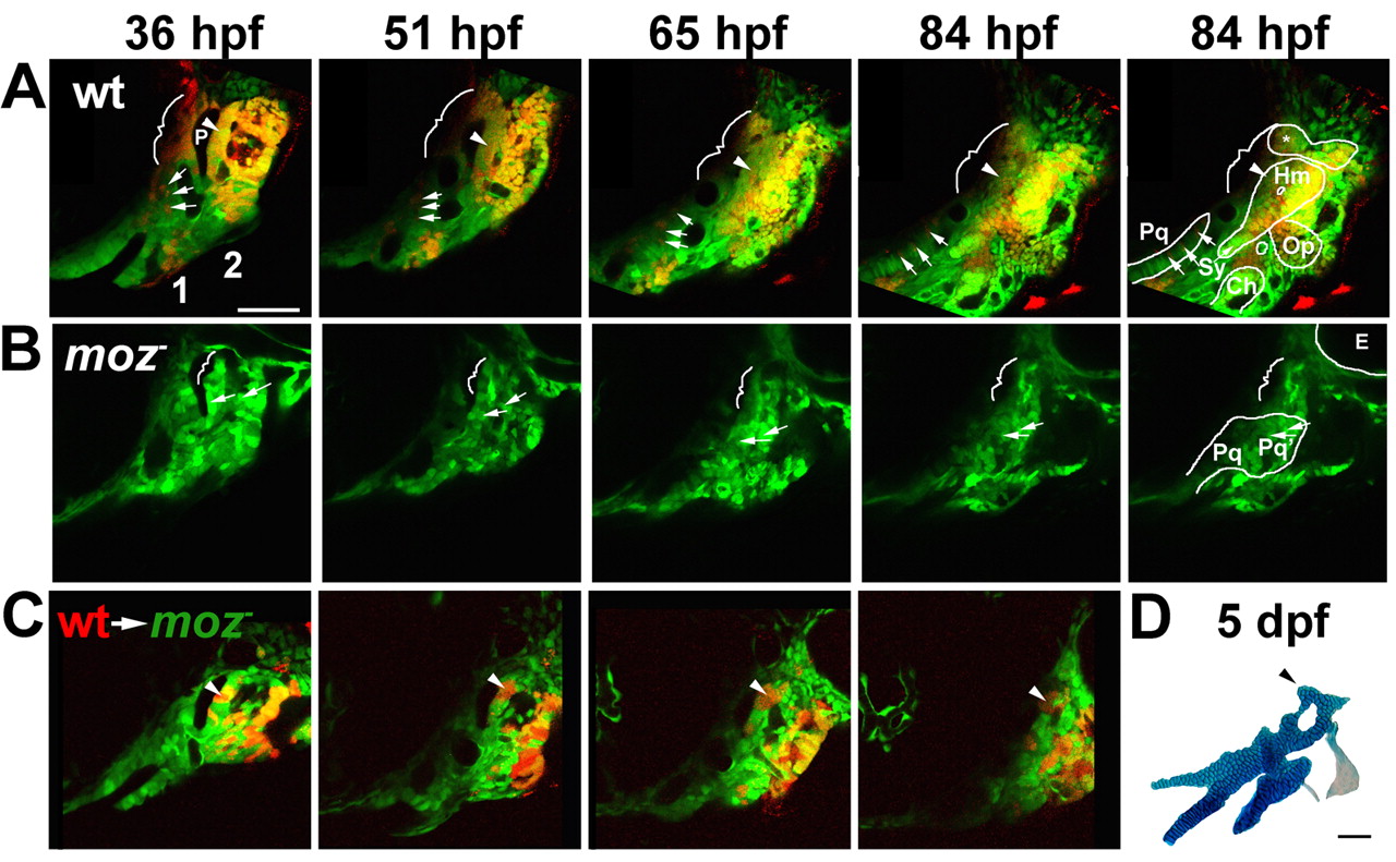
Fig. 6 Moz is required for the growth and chondrification of dorsal second-segment CNC. (A-C) Progressive frames, as indicated, from time-lapse confocal recordings of first (1) and second (2) pharyngeal segment development in wild-type fli1:GFP (A), moz-; fli1:GFP (B), and moz-; fli1:GFP/wild-type fli1:GFP mosaic (C) animals. Skeletal outlines are shown at 84 hpf. (A) A subset of CNC precursors are labelled with a red dye. In wild type (n=4), dorsal second-segment CNC (arrowheads) adjacent to p1 endoderm (P) form the anterior part of Hm cartilage. Dorsal first-segment CNC (brackets) form either no cartilage or neurocranial cartilage (*). Intermediate first-segment CNC (arrows) contribute to Pq cartilage. (B) In moz-; fli1:GFP animals (n=6), intermediate second-segment CNC contribute to Pq' cartilage (arrows). More dorsal CNC make no cartilage (brackets). In C, small amounts of wild-type CNC precursors (red) were transplanted into moz-; fli1:GFP animals. Wild-type second-segment CNC (arrowheads) adjacent to p1 grow and form a cartilage nodule in the normally cartilage-free dorsal zone of the moz- host. (D) The skeletal preparation of this same animal at 5 dpf. Scale bars: 50 µm. See also Movies 1-3 in the supplementary material.