
|
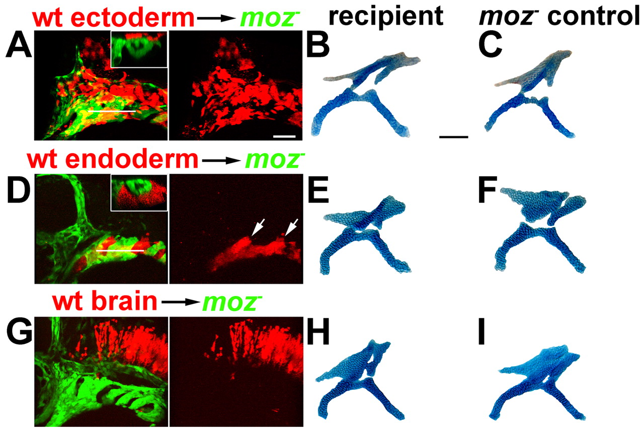
Fig. 3 Moz is not sufficient in the endoderm, surface ectoderm or hindbrain to rescue moz- skeletal development. Wild-type ectoderm (A-C), endoderm (D-F) and hindbrain (G-I) precursors from fli1:GFP animals were unilaterally transplanted into moz-; fli1:GFP hosts at shield stage. Confocal images show that transplants (red) contributed significantly to the surface ectoderm (A), facial endoderm and pouches (arrows in D), and hindbrain (G) at 36 hpf. Insets in A and D are digital longitudinal sections through the level of the white lines and show that transplanted tissue did not express the fli1:GFP CNC marker. Lateral is up. (B,C,E,F,H,I) Facial skeletons of the first two segments from the transplanted animals at 4 dpf. Cartilages from the sides receiving transplants (B,E,H) were indistinguishable from the contralateral control sides (C,F,I). No rescue was seen in 3 ectoderm, 9 endoderm and 12 brain transplants. Scale bars: 50 µm.