
Fig. 6
|
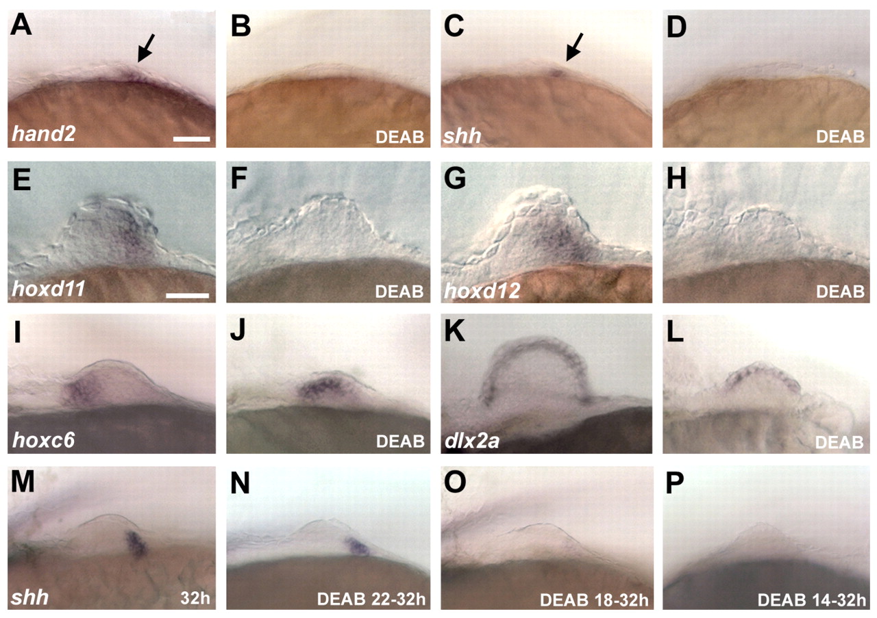
Fig. 6 Expression of marker genes in the mesenchyme and ectoderm of wild-type and pectoral fins. Treatment with 10 µM DEAB, starting from 16 hpf to 28 hpf (A-D), 38 hpf (I,J) and 40 hpf (E-H,K,L); and from differing starting times to 32 hpf (M-P). Anterior is towards the left. (A,B) hand2 is expressed in the medial and posterior mesenchyme of wild type (arrow), but is not expressed in the absence of RA. (C,D) shh is expressed in the posterior mesenchyme (arrow), but is not expressed in the absence of RA. (E-H) hoxd11 and hoxd12 are expressed in the posterior mesenchyme and fail to be induced in the absence of RA. (I,J) hoxc6, which is normally restricted to the anterior mesenchyme, is expanded posteriorly in DEAB-treated embryos. (K,L) dlx2a, a marker of the apical fold, is normally expressed in the absence of RA. (M-P) shh induction is lost upon early inhibition of RA signaling. Scale bars: 50 µm.