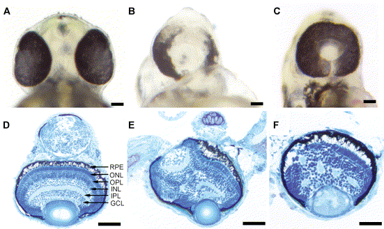
Fig. 9 Overexpression of Pard3 results in cyclopia. Ventral views of the head region of a wild-type embryo at 2 dpf (A), a cyclopic embryo produced by injection of anti-pard3 Morph-1 (B), and a cyclopic embryo generated by injection of wild-type pard3 mRNA (C). In all the pard3 mRNA-injected embryos, the RPE pigmentation was normal. Overexpression of the 150-kDa Pard3 isoform resulted in cyclopic embryos that had normal retinal lamination 95% of the time and disrupted lamination 5% of the time (Panels D and E, respectively). Overexpression of the pard3 mRNA encoding the 180-kDa Pard3 isoform also generated a cyclopic phenotype with 54% of the retinas possessing disrupted lamination (F). The embryos injected with pard3 mRNA also exhibited pericardial edema and curvature of the body axis (data not shown), which may represent nonspecific effects of the mRNA injections as injection of in vitro-transcribed GFP mRNA produced similar frequencies of these phenotypes as pard3-injected embryos. RPE, retinal pigmented epithelium; ONL, outer nuclear layer; OPL, outer plexiform layer; INL, inner nuclear layer; IPL, inner plexiform layer; GCL, ganglion cell layer. Scale bars indicate 100 μm.
Reprinted from Developmental Biology, 269(1), Wei, X., Cheng, Y., Luo, Y., Shi, X., Nelson, S., and Hyde, D.R., The zebrafish Pard3 ortholog is required for separation of the eye fields and retinal lamination, 286-301, Copyright (2004) with permission from Elsevier. Full text @ Dev. Biol.

