
|
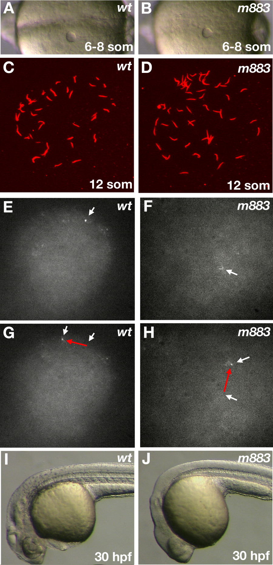
Fig. 6 Kupffer's vesicle in atp1a1a.1 mutant embryos resembles that of wild-type embryos in structure and function. A,B: Morphology of the Kupffer's vesicle at 6-8-somite stage reveals no defects in atp1a1a.1 mutant embryos compared to wild-type. C,D: Detection of monocilia in KV by anti-acetylated tubulin immunohistochemistry. The pictures show flat projections of confocal image stacks. Cilia in m883 mutant embryos appear normal. E-H: Life imaging of monocilia induced fluid flow in embryos injected with 100-nm diameter fluorescent beads. White arrows point at the position of fluorescent beads at successive time points. The red arrows indicate the anticlockwise moving beads in a wild-type embryo (E,G), and in a m883 mutant embryo (F,H). These embryos were allowed to develop and the morphological phenotypes documented (I, J). At 30 hpf, the embryo (I) from E,G can be identified as wild-type, while embryo (J) from F,H reveals a mutant phenotype. Wild-type (A,C,E,G,I), and m883 mutant embryos (B,D,F,H,J). Ventral view, anterior to the right (A,B). Lateral view anterior to the left (I,J).