
|
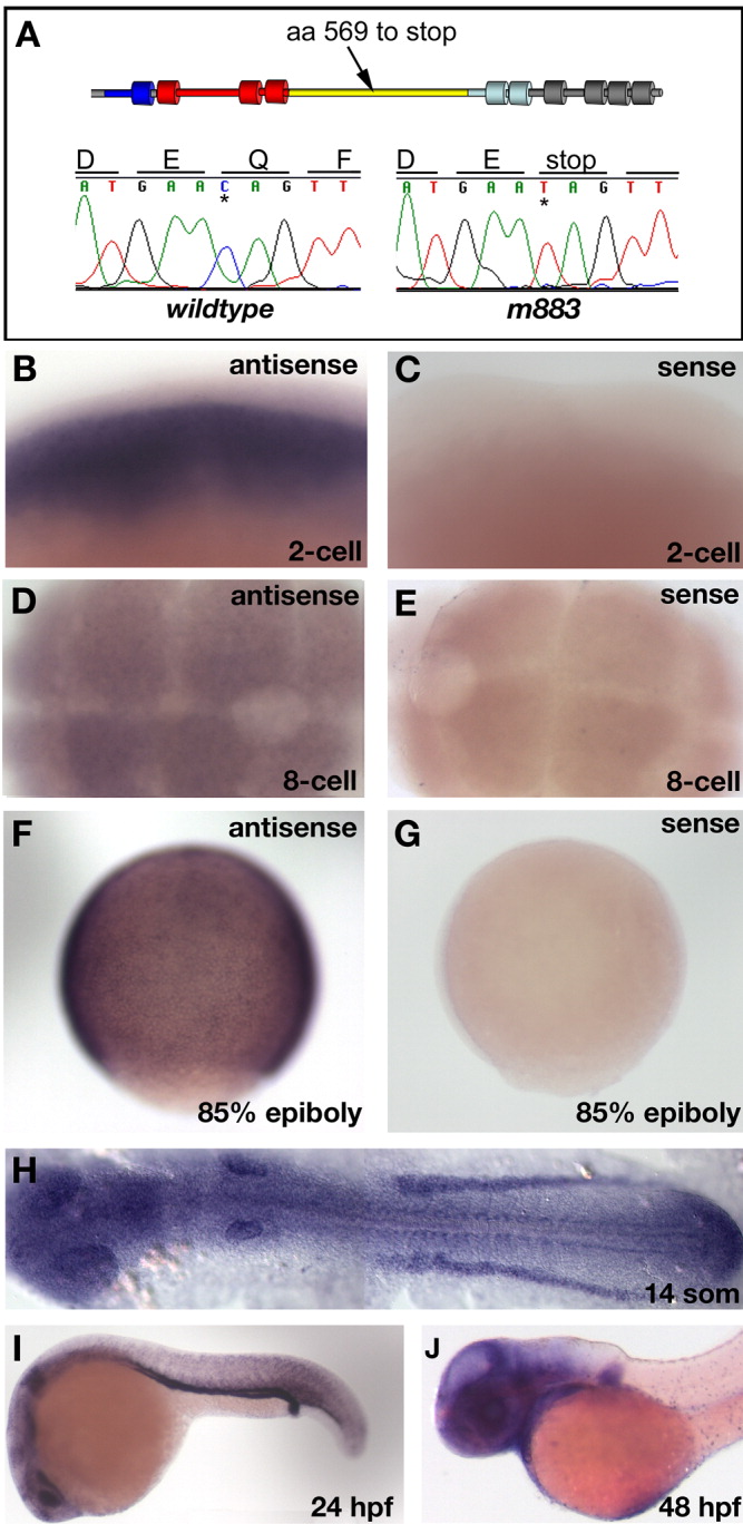
Fig. 3 The m883 mutation truncates the NaK-ATPase alpha1a.1 protein in the hydrolase domain. A: The chromatogram reveals that in the m883 allele, a C to T transition introduced a stop at codon 569, which truncates the NaK-ATPase alpha1a.1 protein in the hydrolase domain. The schematic representation reveals that the stop codon is in the cytoplasmic hydrolase domain (yellow). The thick cylinders represent the transmembrane domains. Dark blue indicates the N-terminal cation transporting ATPase domain, grey indicates the C-terminal cation transporting ATPase domain, red indicates the E1-E2-ATPase domain, light blue indicates the binding domain for the beta subunit. B-J: Whole mount in situ hybridization with an atp1a1a.1-specific probe reveals transcript at the 2-cell stage (B,C) and 8-16-cell stage (D,E). After MBT, the time of onset of zygotic expression, expression remains ubiquitous (85% epiboly in F,G). For all early stages. sense mRNA probes were used as controls (C,E,G). The expression of atp1a1a.1 continues to be ubiquitous during early somitogenesis stages. However, during late somitogenesis (H, 14-somite stage), the expression becomes enhanced in the intermediate mesoderm and in the adaxial mesoderm as well as the ear placode and eye field. At around the 20-somite stage (I, 24 hpf) and later, the expression is most pronounced in the pronephric duct, intermediate mesoderm, ear, eye, brain, and heart. At later stages, 48 hpf (J), the expression is reduced in the pronephric duct and nephric primordium but still remains strong in the brain, eyes, heart, and mucous cells.