
|
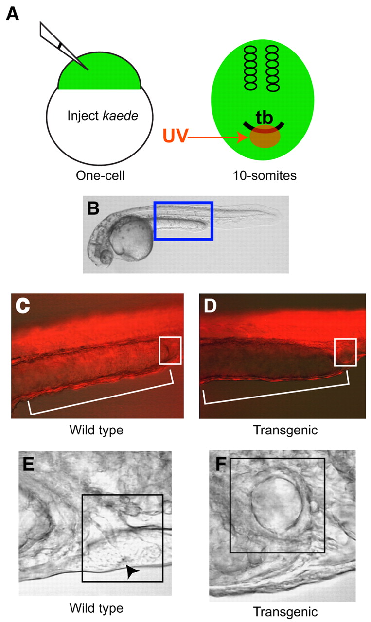
Fig. 2 Lineage labeling of cells ventral to the tail bud reveals severe deficiencies in ventral tissues of transgenic embryos. (A) Scheme for the lineage labeling of extreme ventral cells. (B) Illustration of the labeled region depicted in C and D. Note the decreased number of red fluorescent cells in the ventral yolk extension (bracketed) and cloacal (boxed) region in D compared with in C. (E,F) Visualization of 24 hpf embryos with Nomarski optics revealed severe defects in cloaca formation in transgenic embryos (F) compared with wild-type siblings (E). Arrowhead in (E) depicts the cloacal aperture, and boxed regions in (E) and (F) highlight the pronephric terminus.