
|
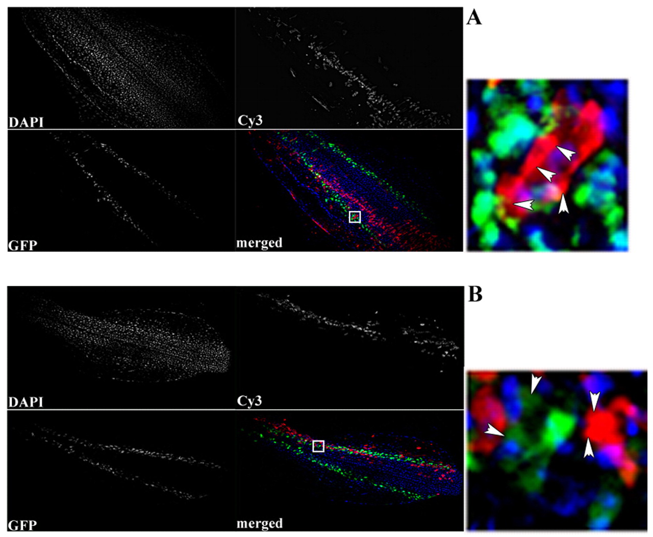
Fig. 7 Excess Bmp4 in lateral mesoderm restricts the number of neighboring hematopoietic cells. Donor cells were derived from embryos that had been injected either with rhodamine-conjugated dextran alone (A) or also with RNA encoding Bmp4 (B). Cells were transplanted into developing embryos that are transgenic for the gata1:gfp reporter. A and B show representative chimeric embryos that contain similar numbers of transplanted (red) cells localized to lateral mesoderm. Embryos were also stained with Hoechst (blue) to distinguish each cell (whether red, green or unlabeled). For each red cell in lateral mesoderm, the number of GFP+ hematopoietic cells in the immediate neighborhood was determined. When donor cells express excess Bmp4, there is a decreased ratio of green (host hematopoietic) to red (donor) cells (data compiled in Table 2). A and B illustrate a single representative embryo for each case, showing the three channels (DAPI, Cy3 and GFP) and a merge, with a small insert indicated in the merged panel on the right. In these magnified panels, the presence of four red cells are indicated by arrowheads. In B, they are associated with a single green (Gata1+) cell, whereas multiple green cells are present in A. Views are flat-mounted dorsal, anterior towards the left.