
|
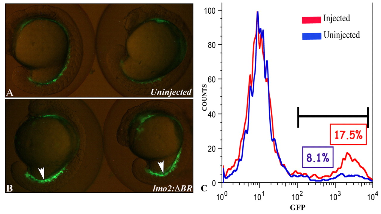
Fig. 4 Expression of a dominant-negative BMP receptor in lateral mesoderm results in enhanced hematopoiesis. Two representative embryos are shown in A and B; views are lateral, anterior towards the top, with embryos still in their chorions. (A) Control embryos from the gata1:gfp transgenic line demonstrate the normal expression pattern of GFP in the ICM. (B) Embryos from the gata1:gfp transgenic line that had been injected at the one-cell stage with the I(lmo2δBR)I transgene and meganuclease. GFP expression is substantially increased in the ICM, compared with control embryos (arrowheads). (C) Dissociated cells were collected at the 16- to 17-somite stage from batches of control and transient transgenic embryos and analyzed by FACS to score quantitatively the numbers of GFP+ cells. Shown are results from one representative experiment, although the data were comparable in three independent experiments. Compared with control embryos (blue) the I(lmo2δBR)I transgenic embryos (red) show more than double the normal amount of GFP+ hematopoietic cells.