
|
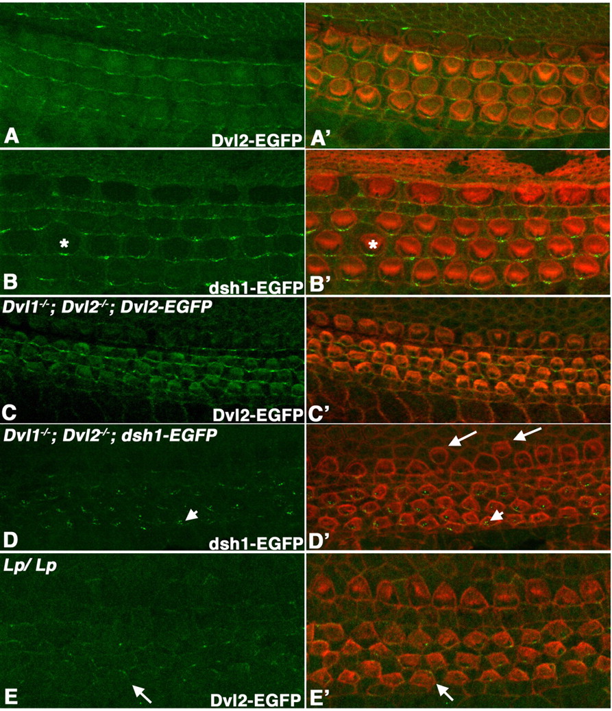
Fig. 6 Localization of Dvl2-EGFP and dsh1-EGFP in the organ of Corti. (A-E) Confocal scanning at the apical surface of hair cells in whole-mount organ of Corti at E18.5, showing the native Dvl2-EGFP (A,C,E) or dsh1-EGFP (B,D) signal. (A'-E') are the overlay of Dvl2-GFP (green, A',C',E') or dsh1-EGFP (green, B',D') and hair cell membrane and stereociliary bundles (red) outlined with phalloidin staining. At E18.5 in an otherwise wild-type background, dsh1-EGFP localization along the distal membrane appeared to be more punctate (asterisk in B and B') when compared with wild-type Dvl2-EGFP (A,A'). By contrast, in Dvl1-/-; Dvl2-/- background at E16.5, dsh1-EGFP localization on the membrane was mostly lost and accumulation in the cytoplasm was observed (arrowheads in D,D'). Dvl1-/-; Dvl2-/-; dsh1-EGFP mutants also display mis-alignment of inner hair cells (arrows in D'). Although wild-type Dvl2-EGFP could maintain an even distribution along the distal membrane in Dvl1-/-; Dvl2-/- background at E16.5 (C,C'), its membrane localization was reduced and no longer restricted to the distal side (arrows in E,E') in Lp/Lp mutants at this stage.