
|
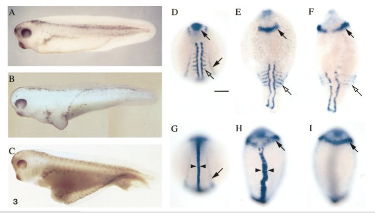
Fig. 3 Dorsalizing activity of zebrafish Chordin in Xenopus and zebrafish embryos. (A) Uninjected Xenopus embryo (stage 37/38); (B) Xenopus embryo coinjected with 10 nl of zebrafish chordin and GFP RNA (20 ng/μl) on the ventral side at the two-cell stage; (C) Xenopus embryo coinjected ventrally at the one-cell stage with 5 nl of zebrafish chordin and GFP RNA (20 ng/μl). (D – I) In situ hybridization using pax2 and myoD probes (D – F) or pax2 and ntl probes (G – I) of uninjected zebrafish embryos (D and G) or embryos injected with chordin RNA (E, F, H, and I) at the one- to four-cell stage during segmentation. All injected embryos shown correspond to class d of Table 2. (E, F) Severely dorsalized embryos were characterized by their elongated shape, laterally expanded domain of pax2 expression at the midbrain – hindbrain junction (arrow), and ventrally extending somites (open arrows). (H) In some injected embryos, the ntl expression domain (arrowheads) in the notochord was widened relative to controls (G). D – H are dorsal views. (I) A ventral view of the same embryo as H shows the expansion of pax2 expression in the brain around the entire circumference of the embryo (arrow). Pronephric duct expression of pax2 (arrow, G) was often missing in injected embryos.
Reprinted from Developmental Biology, 192, Miller-Bertoglio, V.E., Fisher, S., Sanchez, A., Mullins, M.C., and Halpern, M., Differential regulation of chordin expression domains in mutant zebrafish, 537-550, Copyright (1997) with permission from Elsevier. Full text @ Dev. Biol.