
|
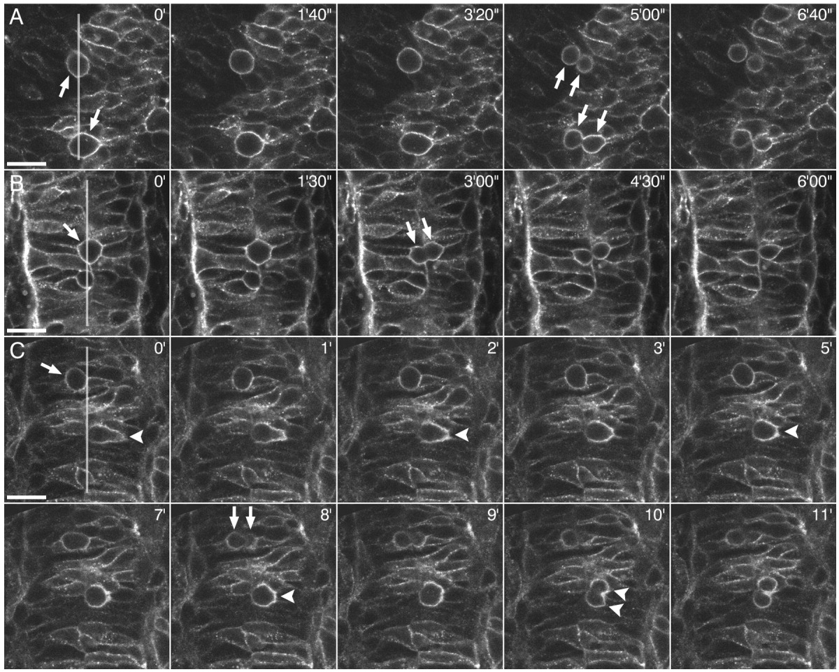
Fig. 4 A-C: Sequences of time-lapse frames of mitotic cells of embryos injected with numb(PTBL PRRL):egfp mRNA. The transparent grey line marks the midline. All photographs are dorsal views. Anterior is towards the top. Scale bar = 20 μm. A: Two mitotic cells in the neural plate of a 5- to 6-somite embryo (arrows in 0'). Cell divisions are planar at this stage. Numb:EGFP is distributed ubiquitously around the cell cortex and segregated to both daughter cells upon cell division (arrows in 5'00"). B: A mitotic cell in the neural rod of a ~16-hpf-old embryo (arrow in 0'). Cells in this stage divide perpendicularly to the midline, due to a 90° rotation of the mitotic spindle. As in the neural plate stage, Numb:EGFP is localized ubiquitously around the cell cortex and, therefore, segregated to both daughter cells (arrows in 3'00"). C: Two mitotic cells during the transition from neural rod to neural tube in a ~17-hpf-old embryo. Whereas one cell (arrow in 0') still divides perpendicular to the plane of the epithelium with Numb:EGFP being ubiquitously localized (arrows in 8'), the other cell (arrowhead in 0') already divides parallel to plane of the epithelium, as is typical for all cell divisions in the neural tube (arrowheads in 10'). Note that this cell starts to localize Numb:EGFP to the basolateral cell cortex when it rounds up at the midline for mitosis (frames 0'- 8'). See also Supplemental Material for the corresponding Supplementary Movies 1 to 3.