
|
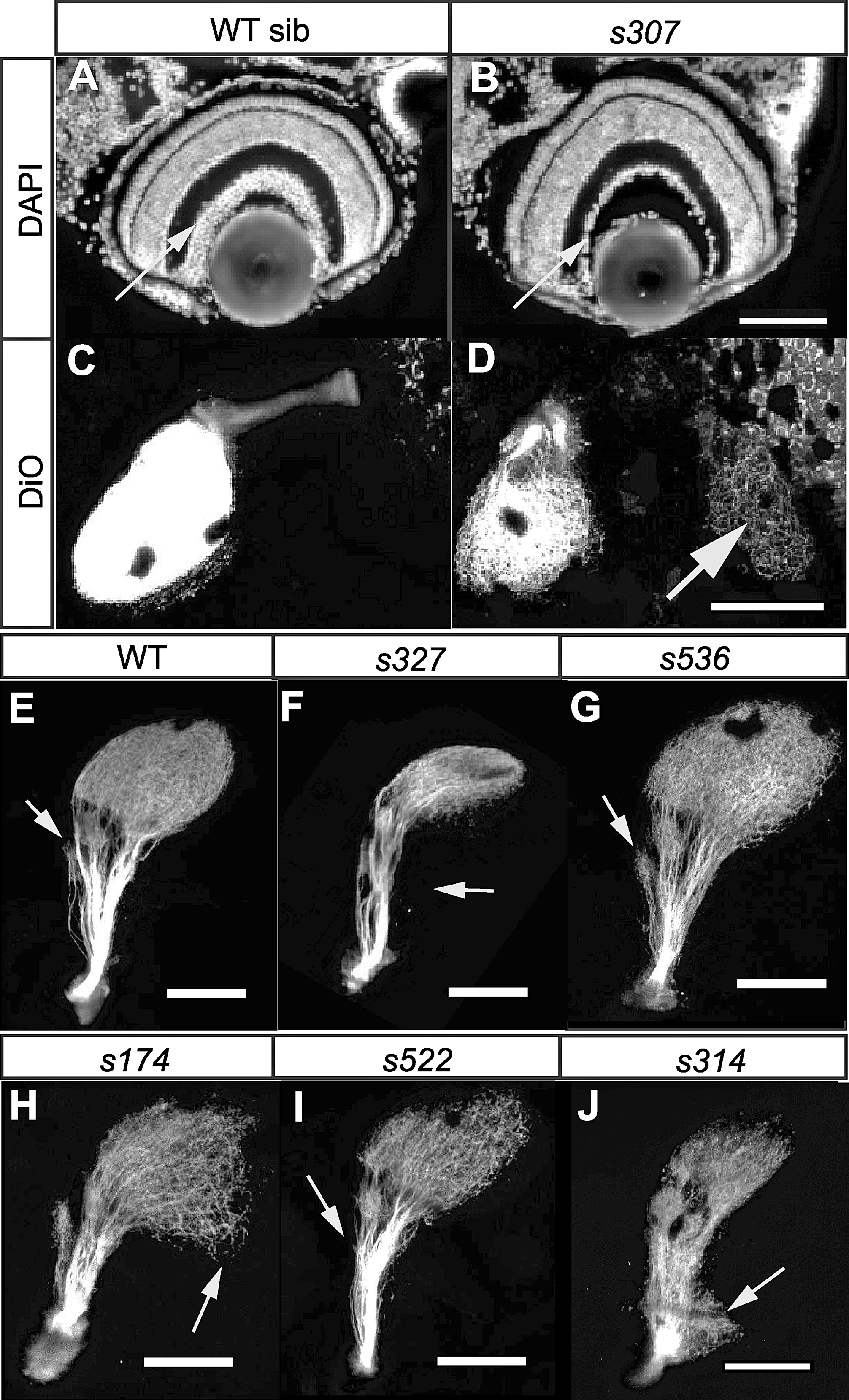
Fig. 7 Examples of Retinofugal Projection Mutants. (A and B) Sections of WT and bojs307 retina stained with DAPI. The mutant retina has a thinner RGC layer (arrow). (C and D) Dorsal views of RGC axons from the right eye of a WT and a bojs307 mutant labeled with DiO, showing mutant axons in the ipsilateral tectum (arrow). To show that there is no ipsilateral projection in WT, the image is overexposed. (E-J) Lateral views of RGC axons labeled with DiO after removal of the eye. Anterior is to the left, dorsal to the top. In WT, the tectum and other retinorecipient areas are clearly visible (E). The arrow indicates AF-4. In darls327, the ventral branch of the optic tract is missing (arrow), and only dorsal tectum is innervated (F). In walks536, innervation of AF-4 (arrow) is disorderly (G). In exas174, the posterior tectum (arrow) appears to be incompletely innervated, while AF-4 is larger than in WT (H). In misss522, AF-4 (arrow) is reduced in size (I). In michs314, there is an ectopic arborization (arrow) at the root of the optic tract (J). Scale bars are 100 μm.