
|
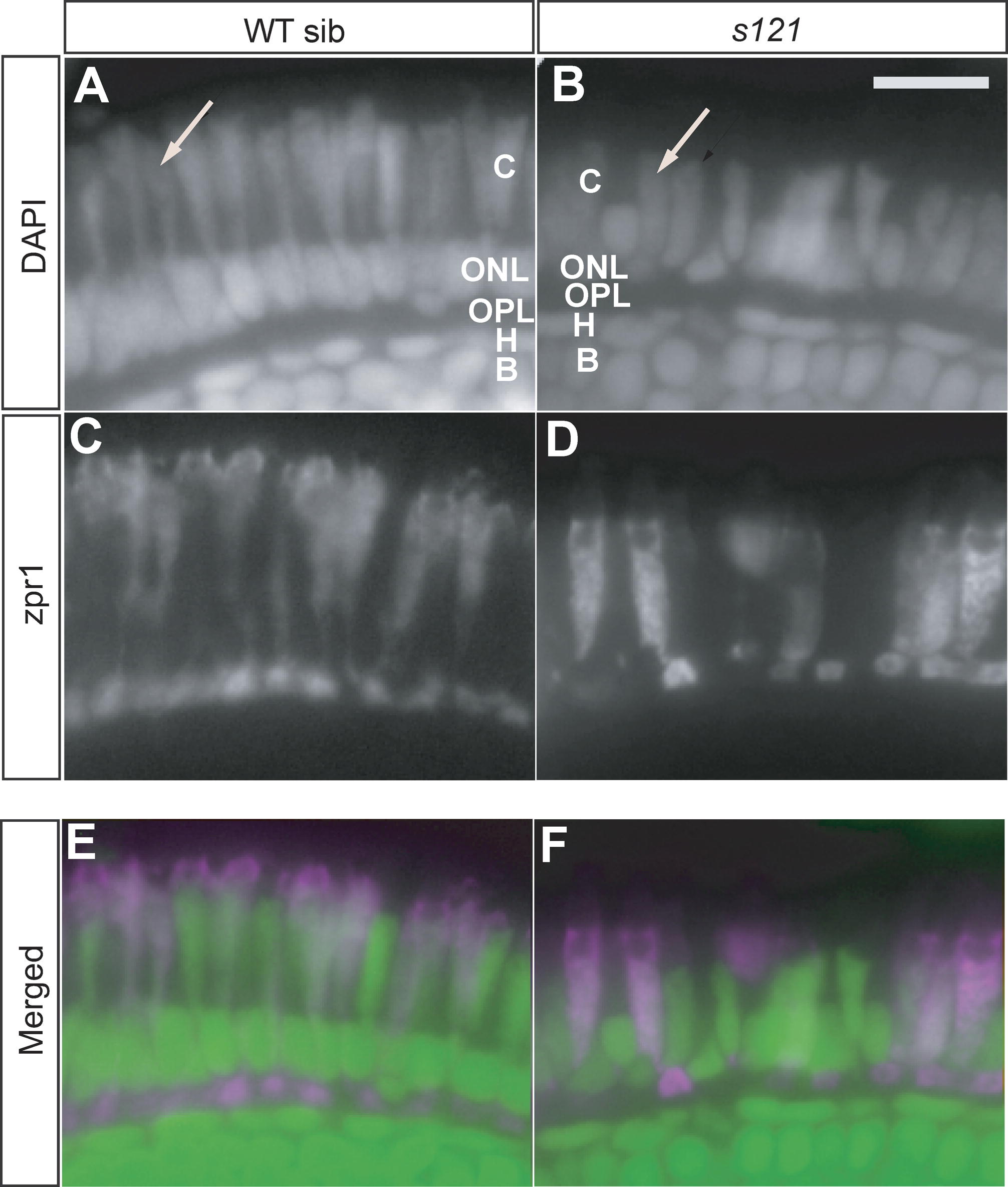
Fig. 3 Example of a Mutant with Abnormal Morphology of Cone Photoreceptors. Photoreceptors in a retinal section stained with DAPI (A and B) and a marker for double cones, zpr1 (C and D) at 7 dpf in WT larva (A, C, and E) and yois121 mutant retina (B, D, and F). Merged images of DAPI (in green) and zpr1 (in magenta) are also shown (E and F). Both zpr1-positive and zpr1-negative cone photoreceptors in the mutant are "stumpy" when compared to those in the control retina (arrows). B, bipolar cells; C, cone photoreceptor cells; H, horizontal cells; ONL, outer nuclear layer; OPL, outer plexiform layer. Scale bar is 10 μm.