
Fig. 4
- ID
- ZDB-IMAGE-060307-4
- Publication
- Sakai et al., 2006 - Semaphorin 3d guides laterality of retinal ganglion cell projections in zebrafish
- All Figures
- Figures for Sakai et al., 2006
|
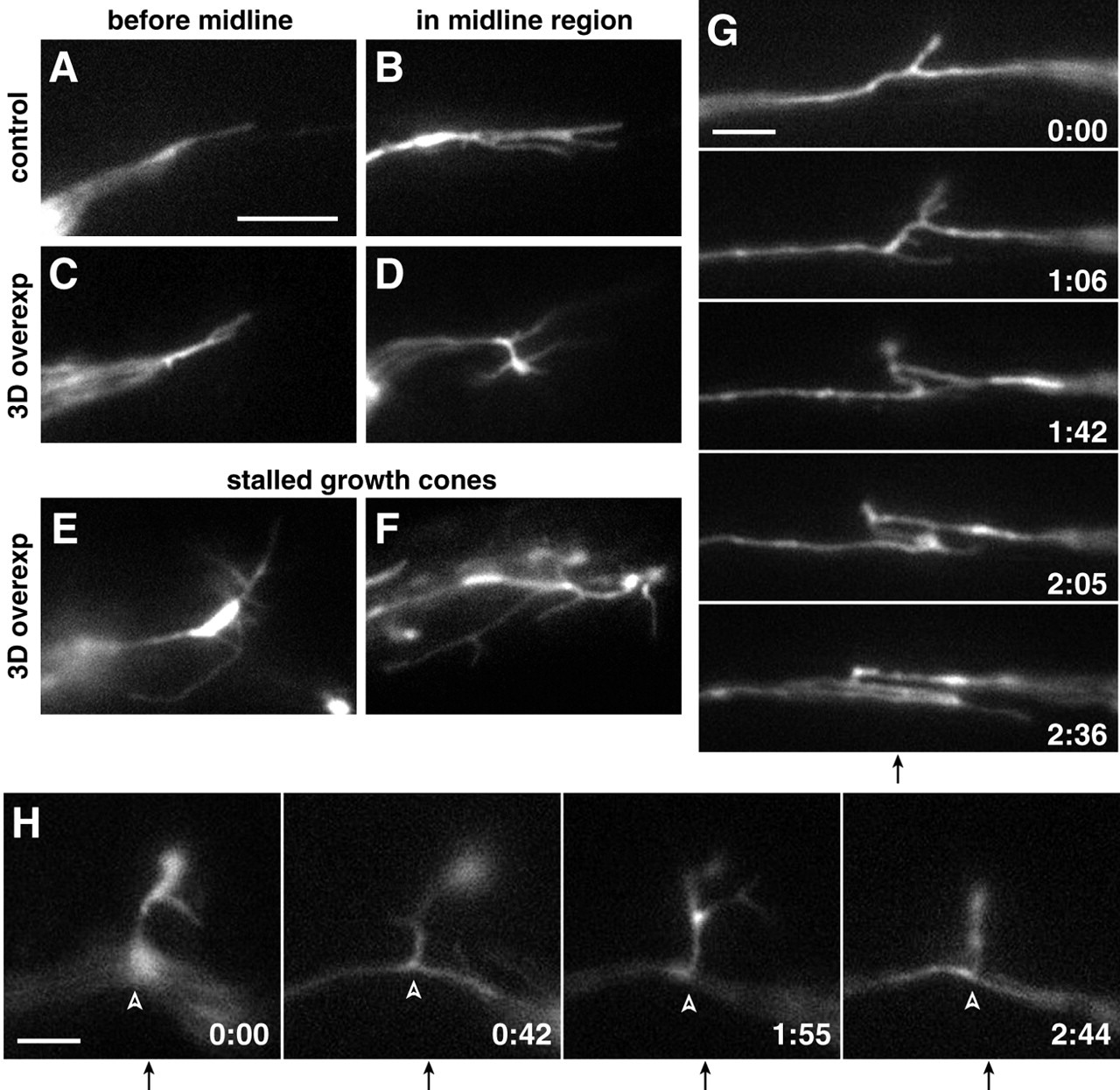
Fig. 4 Live imaging reveals increased growth cone complexity and axonal dynamics following ubiquitous Sema3d expression. (A-D) Single frames from timelapse movies show representative RGC growth cone morphology in wild-type (A,B) and Sema3d-overexpressing (C,D) embryos before and at the midline. Sema3d overexpression did not affect morphology before growth cones reached the midline but increased complexity in the midline region. (E,F) Stalled growth cones in Sema3d-overexpressing embryos showed extremely complex morphology. (G,H) Frames taken from timelapse movies of Sema3d-overexpressing embryos show examples of (G) dynamic axonal movements and (H) an interstitial projection (arrowheads) from the midline. Times are reported in hours:minutes; midlines are indicated by arrows below frame sequences. Anterior is up in all frames. Scale bars: 10 μm in A-F; 5 μm in G,H.