
|
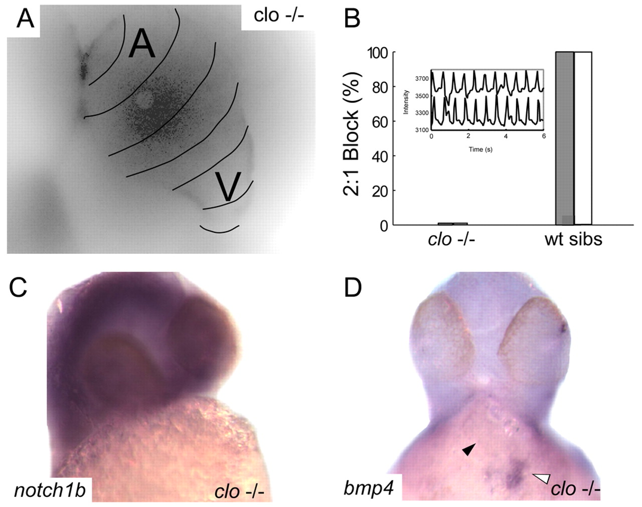
Fig. 3 Endocardial signaling is required for the development of AV conduction tissue. (A) Calcium activation map of a single representative cardiac cycle in cloche mutant embryos at 48 hpf. Isochronal lines (black) obtained by fluorescence microscopy are superimposed on a maximum intensity projection of the same heart and demonstrate the failure of AV conduction tissue development in the absence of endocardium. (B) Lack of AV conduction block in cloche embryos compared with wild-type siblings is demonstrated by atrial pacing (white bars) and terfenadine exposure (gray bars). Inset of contemporaneous recordings of atrial and ventricular contraction using intensity-time plots from regions over the respective chambers in a terfenadine treated cloche embryo demonstrating 1:1 AV conduction. (C,D). In situ expression patterns of notch1b (C) and bmp4 (D) in cloche embryos at 48 hpf. Despite overstaining in the brain there is no evidence of notch1b signal in the heart (C). In D, the black arrowhead indicates the location of the AV ring; the white arrowhead denotes intense expression of bmp4 at the sinus venosus.