
|
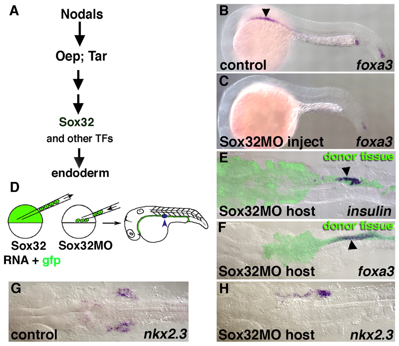
Fig. 4 A transplantation technique using SOX32 to target reagents to specific germ layers. (A) Genetic pathway leading to endoderm formation. SOX32 acts downstream of Nodals, their receptors (TAR) and receptor co-factors (OEP) in endodermal specification. (B) Lateral view of foxa3 expression in a 24 hpf control embryo. Endodermal foxa3 expression extends from slightly anterior of the AP level of somite one (arrowhead) towards the posterior, there is also expression in the posterior tip of the notochord. (C) foxa3 is not expressed in the endoderm of SOX32-MO injected embryos, while notochordal expression in the tail is retained. (D) Schematic of the transplantation technique using sox32 to target donor cells to the endoderm. Donor cells from embryos co-injected with SOX32 and GFP mRNA are placed along the blastoderm margin of a SOX32-MO injected host. Following gastrulation, all endoderm is derived from donor cells, which distribute along the ventral AP axis and exhibit regionalized expression of an array of endoderm-specific markers, including insulin (blue arrowhead). (E-H) Transplanted specimens in dorsal view at 24 hpf. (E) insulin expression (arrowhead) in donor-derived endoderm (green). (F) Normal 24 hpf foxa3 expression (arrowhead) in donor-derived endoderm (green). (G) Normal pharyngeal nkx2.3 expression. (H) nkx2.3 expression in donor-derived endoderm; donor tissue has contributed to only the right side in this example but AP patterning is normal.