
Image
|
Figure Caption
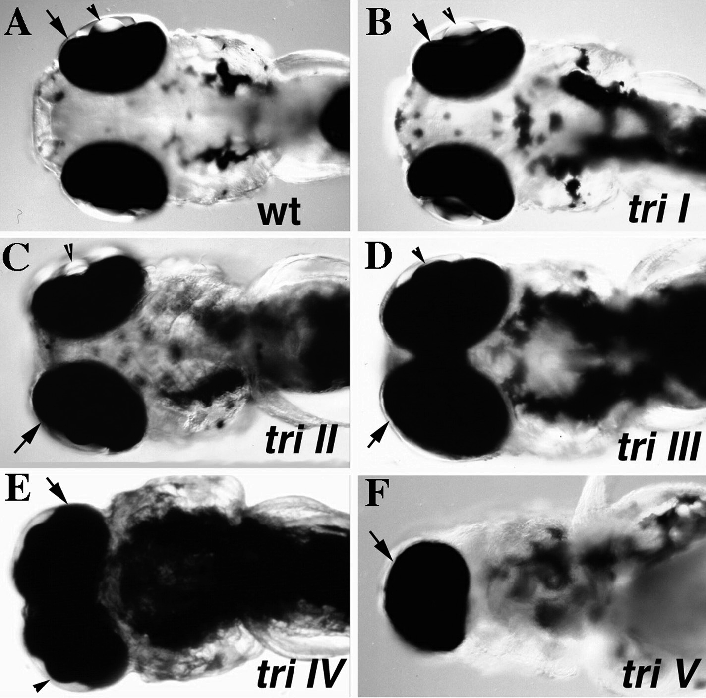
Fig. 1 Degrees of cyclopia in trim209 mutants. Micrographs of live embryos at 3 dpf, ventral view of head region. Five classes of the cyclopia phenotype can be distinguished in trim209 mutants. In class I mutants (B) the eye spacing is comparable to that observed in a wild-type sibling (A). In mutants of class II (C) the eye spacing is decreased. (D) In mutants of class III eyes are marginally fused. (E) The eyes are completely fused in mutants of class IV. (F) One cyclopic eye is observed in class V mutants. Retina covered by pigment epithelium (arrow).
Acknowledgments
This image is the copyrighted work of the attributed author or publisher, and
ZFIN has permission only to display this image to its users.
Additional permissions should be obtained from the applicable author or publisher of the image.
Reprinted from Developmental Biology, volume 203, Marlow, F., Zwartkruis, F., Malicki, J., Neuhauss, S.C.F., Abbas, L., Weaver, M., Driever, W., and Solnica-Krezel, L., Functional interactions of genes mediating convergent extension, knypek and trilobite, during the partitioning of the eye primordium in zebrafish, 382-399, Copyright 1998, with permission from Elsevier.
Reprinted from Developmental Biology, 203, Marlow, F., Zwartkruis, F., Malicki, J., Neuhauss, S.C.F., Abbas, L., Weaver, M., Driever, W., and Solnica-Krezel, L., Functional interactions of genes mediating convergent extension, knypek and trilobite, during the partitioning of the eye primordium in zebrafish, 382-399, Copyright (1998) with permission from Elsevier. Full text @ Dev. Biol.