
|
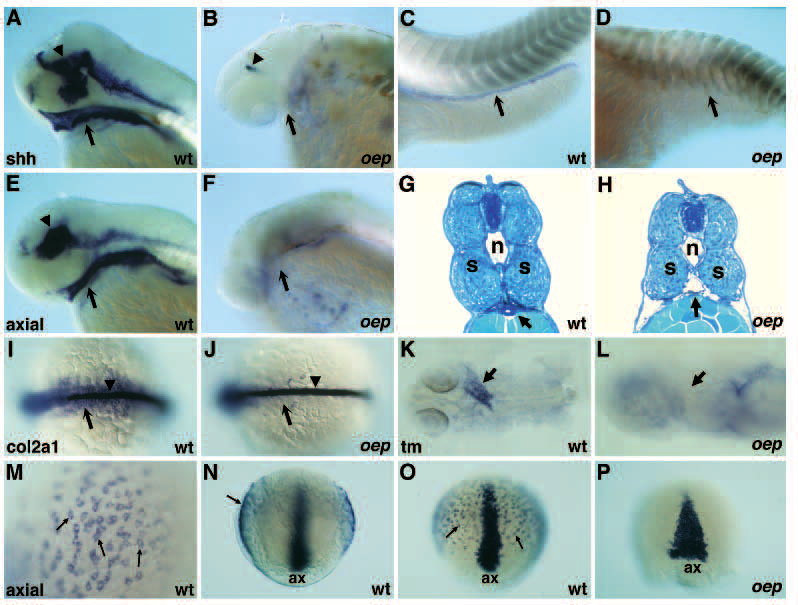
Fig. 6 Endoderm formation is impaired in oep mutant embryos. (A-D) Expression of sonic hedgehog (shh) in wild-type (A,C) and oep mutant (B,D) embryos at 51 hpf. Expression of shh in the pharyngeal endoderm (arrow in A,B) and gut (arrow in C,D) is normal in wild-type but not in oep mutant embryos. Expression in the brain region of oep mutants (arrowhead in B) is strongly reduced as compared to wild-type (arrowhead in A). (E,F) Expression of axial in wild-type (E) and oep mutant (F) embryos at 51 hpf. Expression of axial in the pharyngeal endoderm (arrow in E,F) is normal in wild-type but not in oep mutant embryos. Expression in the brain of oep mutants is also severely affected as compared to wild-type (arrowhead in E). Sagittal cross-section (5 µm) of the trunk region of wild-type (G) and oep mutant (H) embryo at 53 hpf. Arrow indicates the location of the gut in wild-type (G) embryo. Note the lack of tissue and the gaping hole in this region of oep mutants (arrow in H). n, notochord; s, somites. (I,J) Expression of the type II collagen gene col2a1 in wild-type (I) and oep mutant (J) embryos at the 12-somites stage. Note the normal expression in the notochord (arrowhead), but severe reduction in the endoderm (arrow) of oep mutants (J). Dorsal view, anterior is to the left. (K,L) Expression of a-tropomyosin (tm) in the heart region (arrow) of wild-type (K) and oep mutant (L) embryos at 28 hpf. Note the severe reduction of tm in oep mutants. (M-P) Expression of axial in wild-type (M-O) and oep mutant embryos (P) at 80% epiboly. (M) High magnification view of axial expressing cells (arrows) located in the hypoblast. (N) Optical cross section reveals direct juxtaposition of axial-expressing cells to yolk (arrow), ax, axial expression in axial mesoderm. (O) Axial expression in axial mesoderm (ax) and presumptive endoderm (arrows) in wild-type embryos. (P) Loss of axial expression in the presumptive endoderm of oep mutants; ax, axial expression in the axial mesoderm of oep mutant embryos. Axial expression is laterally expanded as a result of reduced convergence and extension in oep mutants.