
|
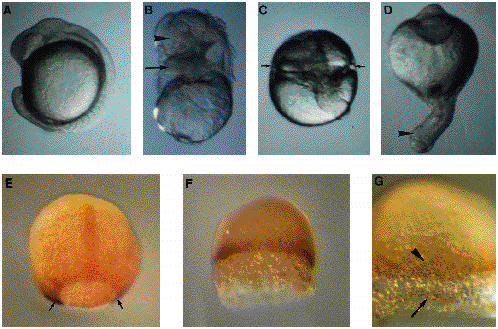
Fig. 6 Treatments of blastula stage embryos with LiCl. (A-D) Micrographs of living embryos at 16 h. (A). Side view of a control embryo at the 14-somite stage. (B-D) Phenotypes of embryos treated at the early blastula (32-cell) stage with 0.3 M LiCl for 30 minutes. (B) Embryo with structures located atop yolk, morphologically identified to brain (arrowhead) and heart (arrow). (C) Embryo with a radial constriction (arrows) possibly indicating heart development. A remnant of axis is visible. (D) Radial proboscis embryo with an external proboscis indicated by an arrowhead. (E-G) Immunodetection of the Ntl protein and whole-mount in situ using a eve1 probe. (F,G). Embryos were treated as in B-D and fixed at 9 h. (E) The positive nuclei for Ntl are visible in notochord precursors and around the margin. The ventral eve1 purple signal (arrows) is hardly visible in this embryo viewed from the dorsal side, animal pole at the top. (F,G) Embryo treated with LiCl. Detail in G. Animal pole up. Yolk was accidentally removed during staining experiments probably as a result of a greater fragility of the embryo. In treated embryos, the external YSL is larger than in untreated embryos. There is no staining for eve1. The Ntl-positive nuclei are located at the margin (arrowhead in G) and in detached cells (arrows in G).