
|
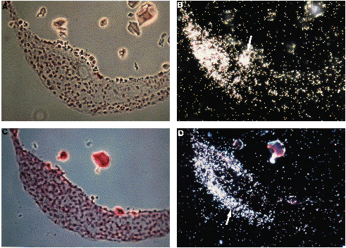
Fig. 5 Expression of eve1 in 10-somite embryos (14 h). (A). Bright-field section of a sagittal section passing through the Kupffer’s vesicle. Yolk has mostly disappeared during the manipulations. (B). Corresponding dark-field view. RNA are located at the tip of the tailbud on the left. A small mass of positive cells (arrow) are located behind the Kupffer’s vesicle (which is negative). (C). Bright-field view of a neighbour section (one section thickness is intercalated between the section shown in A,B and this one). (D). Dark-field view of the region seen in (C). eve1 signal forms an extension in an external cell layer (arrow).