
|
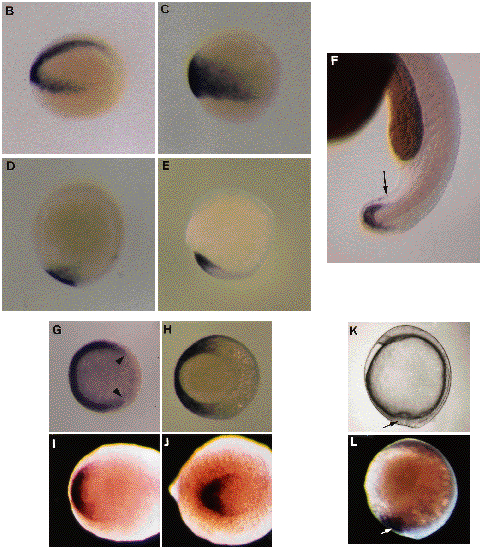
Fig. 4 Localization of eve1 RNA in early embryos. (B-J,L). Whole-mount in situ hybridizations using a eve1 probe. Cells expressing eve1 appear in dark purple after an alkalinephosphatase chromogenic reaction. (B-F). Side views of embryos with the same orientation as in A. (B). 30% epiboly (4.7 h) embryo slightly inclined to see the whole arc of positive cells from the vegetal pole. (C). 60% epiboly (7 h). (D). 100% epiboly (9.5 h). (E). 5-somite embryo (12.5 h). (F). 24h embryo. Positive external cells are located around the extremity of the tail (arrow). (G-J). Views from the animal pole in (G), from the vegetal pole in (H) and from the posterior pole in (I,J). Dorsal side on the right. (G). 30% epiboly stage, the positive signal covering more than 270° of arc (limits indicated by arrowheads). (H). 80% epiboly (8.5 h) stage, positive cells in the ventral half of the gastrula. (I). 1-somite embryo (10.5 h). (J). 10-somite embryo (15 h). (K). Micrograph of a living embryo (9.5 h). Side view. Vegetal pole is at the bottom. The dorsal tail bud is on the right from yolkplug closure. Ventral cells (arrow) converge with their dorsal counterparts. (L). Embryo at the same stage and with the same orientation as in (K). The signal (arrow) forms a plaque ventral to the yolk plug closure and to the dorsal tail bud.