
|
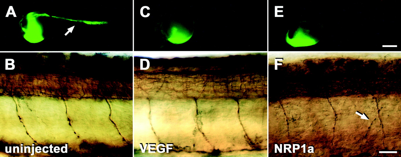
Fig. 6 Differential effects of different morpholinos on blood vessel and on motor axon development in the trunk of 24 hours postfertilization (hpf) embryos. A,C,E: Lateral views of whole embryos subjected to microangiography are shown. Fluorescence of yolk sacs is near the injection site and does not indicate the presence of blood vessels. B,D,F: Mid-trunk levels of embryos labeled with anti-tubulin antibodies subsequent to micro-angiography are shown (rostral is left). A,B: In uninjected embryos, the trunk vasculature (A, arrow) and ventral motor nerves (B) develop normally. C,D: In embryos injected with 1 mM vascular endothelial growth factor (VEGF), morpholino trunk vessels fail to develop (C) but motor nerves grow normally (D). E,F: In embryos injected with 1 mM NRP1a morpholino1, no trunk vessels are labeled (E) and ventral motor nerves grow abnormally (F). Arrow in F indicates a branched ventral motor nerve. Scale bars = 250 μm in E (applies to A,C,E), 25 μm in F (applies to B,D,F).