
Fig. 7
- ID
- ZDB-IMAGE-060119-9
- Publication
- Alioto et al., 2005 - The odorant receptor repertoire of teleost fish
- All Figures
- Figures for Alioto et al., 2005
|
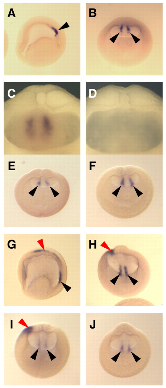
Fig. 7 Bilateral expression of XNR1 is not affected by XCR2 MO injection. (A-C) Normal expression pattern of XNR1 in the posterior archenteron roof at stage 15 as determined by in situ hybridization using uninjected embryos. Asymmetric expression of XNR1 was not observed at stage 15. (C) Posterior archenteron roof of half-dissected embryo in side view. Two stripes of bilateral XNR1 expression were observed. (D) In situ hybridization with an XNR1 sense probe. No signal was observed in the archenteron roof. (E,F) Injection of 20 ng of SC MO (E) or of XCR2 MO (F) into the marginal region of both blastomeres at the two-cell stage. The bilateral expression of XNR1 is not affected at stage 15 in either condition. (G,H) Normal expression pattern of XNR1 at stage 20. The bilateral expression is maintained (black arrowheads) and asymmetric expression appears in the LPM (red arrowhead). (I) Injection of 20 ng of SC MO does not inhibit either bilateral or asymmetric expression of XNR1 at stage 20. (J) Injection of 20 ng of XCR2 MO inhibited the asymmetric expression of XNR1, but not bilateral expression at stage 20. (A,G) Lateral views. The anterior side is to the left. (B,E,F,H-J) Posterior views. The dorsal side is up. The embryo was cleared with benzyl benzoate/benzyl alcohol (A,B,E-J). Black arrowheads indicate the bilateral expression of XNR1; red arrowheads indicate the asymmetric expression of XNR1.