
Fig. 6
- ID
- ZDB-IMAGE-060119-8
- Publication
- Alioto et al., 2005 - The odorant receptor repertoire of teleost fish
- All Figures
- Figures for Alioto et al., 2005
|
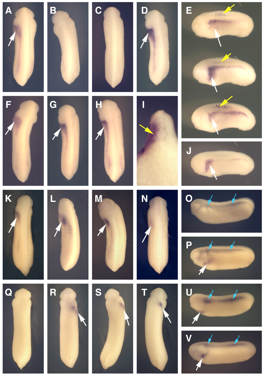
Fig. 6 Left-side injection of XCR2 MO inhibits XNR1 and XATV expression in the lateral plate mesoderm (LPM). (A-N) In situ hybridization of XNR1. White arrows indicate the expression of XNR1 in the LPM. (A) Embryo injected with 10 ng of SC MO into the left side, showing normal XNR1 expression. (B) Injection of 10 ng of XCR2 MO into the left side caused no expression of XNR1 in the LPM. (C) Co-injection of 10 ng of XCR2 MO with 10 pg of pCS-EGFP plasmid into the left side caused no expression of XNR1 in the LPM. (D,E,I) Co-injection of 10 ng of XCR2 MO with 10 pg of pCS-XCR2 plasmid into the left side rescued the expression of XNR1 in the LPM and also caused ectopic XNR1 expression in the somite (yellow arrows). (I) Transverse section of the rescued embryo. Ectopic XNR1 expression in the somite was observed. (F,G) Right-side injection of 10 ng of SC MO (F) or XCR2 MO (G) did not affect XNR1 expression. (H,J) Uninjected control embryo. (K-N) Co-injection of 10 ng of XCR2 MO with 10 pg of XCR1 (K), XCR3S (L), XCR3L (M) or human CFC1 (N) plasmid in the left side rescued the expression of XNR1 in the LPM. (O-V) In situ hybridization of XATV in ventral view. Arrows indicate the expression of XATV in the left LPM (white) and the dorsal midline (blue). (O,Q) Embryo injected with 10 ng of XCR2 MO in the left side inhibited the expression of XATV in the left LPM, but not in the dorsal midline. (P,R) Embryo injected with 10 ng of XCR2 MO in the right side, showing normal XATV expression. (S,U) Co-injection of 10 ng of XCR2 MO with 10 pg of pCS-XCR2 plasmid in the left side rescued the expression of XATV in the left LPM. (T,V) Uninjected control embryo.