
Fig. 5
- ID
- ZDB-IMAGE-060119-7
- Publication
- Alioto et al., 2005 - The odorant receptor repertoire of teleost fish
- All Figures
- Figures for Alioto et al., 2005
|
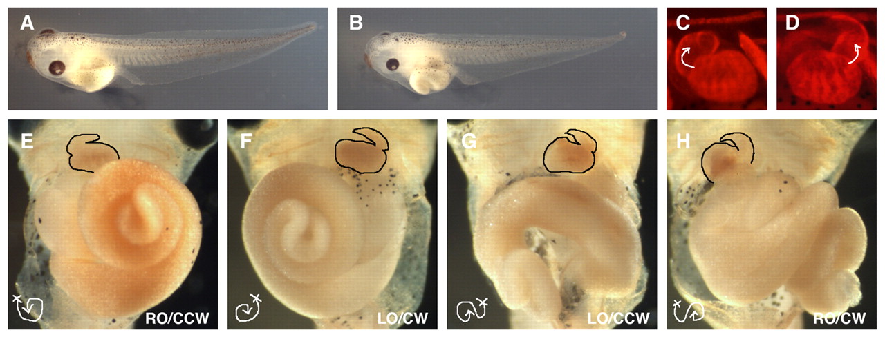
Fig. 5 XCR2 MO disturbs randomization of left-right asymmetric patterning of the heart and gut in stage 45-46 embryos. (A) Embryo injected with 50 ng of a standard control MO (SC MO). (B) Embryo injected with 50 ng of XCR2 MO. (C-H) Embryos injected with MO into the marginal region of both blastomeres at the two-cell stage, immunostained with MF 20 antibody, and the signal detected by fluorescence (C,D) or DAB staining (E-H). (C,E) Embryo injected with 50 ng of SC MO, showing normal left-right asymmetric patterning of the heart (C,E) and gut (right origin and counterclockwise coil, RO/CCW) (E). (D,F-H) Embryos injected with 20 ng of XCR2 MO. (D) Reversed morphology of heart. (F) Embryo has a mirror-imaged heart and gut (the left origin and clockwise coil, LO/CW). (G) Embryo has a normal heart, and left origin and counterclockwise-coiled gut (LO/CCW). (H) Embryo has reversed heart, right origin and clockwise-coiled gut (RO/CW).