
|
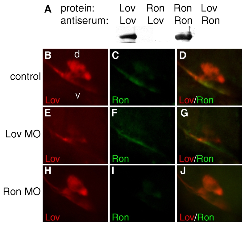
Fig. S2 Specificity of Leftover and Right on antisera. (A) Western blot showing detection of bacterially expressed Lov or Ron protein (1 mg) by anti-Lov (lanes 1, 2) and anti-Ron (lanes 3, 4) rabbit polyclonal antiserum, and the lack of cross-reactivity. (B-J) Embryos injected with 3 ng of lov or ron morpholino (complementary to the 5¢ UTR proximal to the start ATG) were harvested at 3.5 days, at which time Lov and Ron proteins were detected by immunofluorescence of habenular projections to the interpeduncular nucleus. Upon injection of Lov MO, Lov (E) but not Ron immunofluorescence (F) was significantly reduced relative to uninjected control larvae (B,C). Conversely, ron MO injections resulted in reduced Ron immunofluorescence (I) at the IPN, but did not affect Lov labeling (J). All confocal images are lateral views of the dorsal (d) and ventral (v) IPN, as indicated in B.