
|
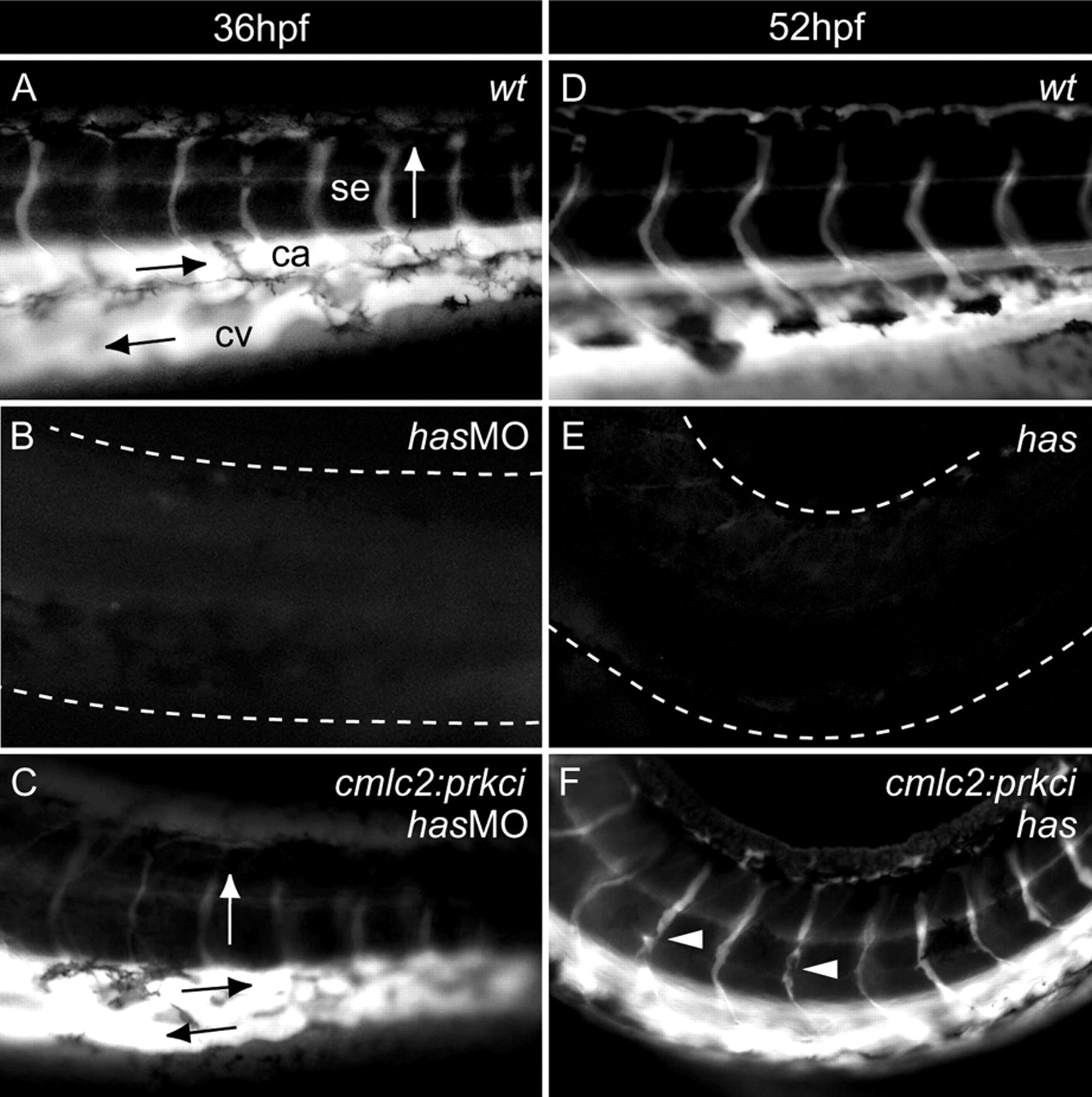
Fig. 2 Has/PRKCi activity within myocardial cells is sufficient for heart function. Microangiograms depicting the peripheral circulation present within the tail region of embryos. The direction of blood flow is indicated by arrows. (A) wild-type embryo at 36 hpf. (B) has/prkci morphant at 36 hpf. (C) Tg(cmlc2:prkci) transgenic animal injected with the has/prkci MO at 36 hpf. (B) By contrast, peripheral circulation is never seen in has/prkci morphants. (D) Wild-type embryo at 52 hpf, with robust circulation. (E) Complete lack of peripheral circulation in a has mutant. (F) At 52 hpf, peripheral circulation is present in has mutants with Tg(cmlc2:prkci) insertion but defects in vessel formation can be observed (arrowheads). The white dotted line indicates the outline of the tail region. ca, caudal aorta; cv, caudal vein; se, segmental vessel.