
|
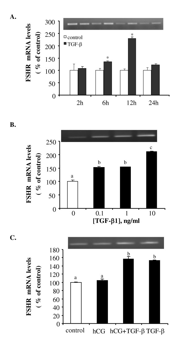
Fig. 4 TGF-β1 stimulates FSHR mRNA expression. (A) Follicles were incubated with control medium or 10 ng/ml of TGF-β1 for 2, 6, 12 and 24 hours. (B) Follicles were treated with different concentrations (0, 0.1,1 and 10 ng/ml) of TGF-β1 for 18 hours. (C) Follicles were treated with medium (control), hCG (100 IU/ml), TGF-β1 (10 ng/ml), or a combination of hCG and TGF-β1 for 18 hours. At the end of each incubation, total RNA was extracted and reverse transcribed. PCR was carried out using primers for FSHR and GAPDH. Each value represents the mean ± SEM of three replicates in one representative RT-PCR. Statistical significance (P < 0.05) is indicated by either an * or a different letter. The insets show the representative ethidium bromide stained gels.