
|
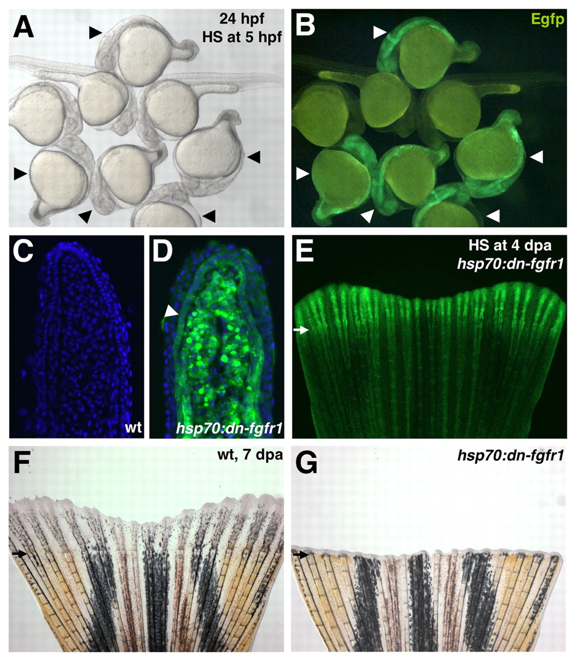
Fig. 5 Transgenic fish that facilitate inducible expression of a dominant-negative Fgfr1 construct. (A,B) hsp70:dn-fgfr1 transgenic and wild-type embryos were raised until sphere stage at 28°C, shifted to 37°C for 1 hour, and returned to 28°C until 28 hpf. Transgenic embryos are truncated and display Egfp fluorescence (arrowheads). (C,D) Section through a wild-type (C) or hsp70:dn-fgfr1 (D) 4 dpa fin regenerate 5 hours after heat induction. Strong Egfp fluorescence is observed in all cells of the transgenic regenerate, including the fgfr1/mkp3/sef/spry4-positive basal epidermal layer (arrowhead). Nuclei are stained with DAPI (blue). (E) Whole-mount image of fin shown in D, demonstrating Egfp fluorescence throughout the fin. The regenerate shows stronger fluorescence than the non-regenerating portion at 4 dpa. This is probably due in part to the scarceness of pigment cells and differentiated bone in the newly formed tissue that might impede fluorescence detection. (F,G) Adult fin regeneration is blocked by daily heat-induction of dn-fgfr1 at 38°C. Wild-type (F) and hsp70:dn-fgfr1 (G) fins are shown at 7 dpa.