
|
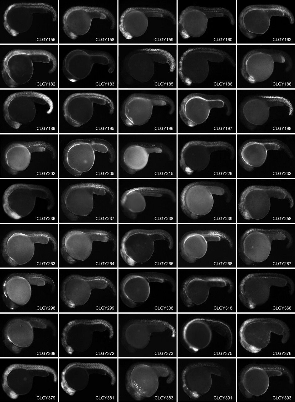
Fig. 2 Stable transgenic lines showing spatially and temporally restricted expression of YFP around 24h post fertilization, anterior is to the left and dorsal to the top. Main domains of expression at 1 dpf (CLGY number is in parentheses): (1) Widespread expression, most pronounced in hypothalamus and posterior somites. (2) Diencephalon, hindbrain and anterior spinal cord. (3) Olfactory placodes. (4) Yolk syncytial layer, ventral caudal mesoderm. (5) Retina, epiphysis and spinal cord. (6) Developing skeletal muscle. (7) Widespread in CNS and muscle. (8) Dorsal telencephalon. (9) Hindbrain and posterior mesenchymal cells. (10) Subset of cells in CNS and notochord. (11) Neural crest and cranial ganglia. (12) Differentiating skeletal muscle and notochord. (14) Widespread, pronounced in telencephalon, retina, hindbrain and most posterior tissues. (16) Tail tip, notochord and skeletal muscle. (18) CNS, hindbrain (19) Dorsal telencephalon and mottled expression in notochord and spinal cord. (20) Hindbrain and spinal chord. (21) Developing skeletal muscle. (22) Telencephalon, olfactory placodes, hindbrain and spinal cord. (24) Widespread expression in CNS and muscle. (30) Hindbrain and spinal cord. (31) Retina and differentiating muscle. (32) Notochord and differentiating muscle. (33) Retina, hindbrain and spinal cord. (35) Widespread expression in CNS and muscle. (38) Retina, mid-hindbrain and hindbrain. (39) Sheath cells of the notochord (41) Retina, telencephalon and hindbrain. (47) Posterior mesenchymal cells. (50) Tail tip. (65) Telencephalon, olfactory placodes, hindbrain and spinal cord. (68) Widespread. (69) Spinal cord, posterior to somite 4/5 boundary. (75) CNS. (77) Spinal cord. (86) Telencephalon, hindbrain and spinal cord. (97) Notochord and differentiating muscle. (101) Telencephalon and differentiating muscle. (102) Differentiating skeletal muscle. (104) Cells in hindbrain and anterior part of spinal cord. (109) Forebrain and spinal cord. (128) Widespread, pronounced in ventral diencephalon and tailbud. (129) Hatching glands. (130) Retina and differentiating muscle. (131) Blood, retina, telencephalon, hindbrain, and muscle. (135) Posterior trunk. (142) Widespread. (143) Skeltal muscle and CNS. (151) Dorsal diencephalon and dorsal mesencephalon. (154) Widespread, strong in retina and telencephalon. (155) Telencephalon, retina, midbrain, rhombomeres, spinal cord. (158) Widespread. (159) Central nervous system and developing muscle. (160) Telencephalon and diencephalon. (182) Widespread in CNS and muscle. (183) Telencephalon. (185) Spinal cord and posterior trunk. (186) Central nervous system. (188) Retina and telencephalon. (189) Widespread. (195) Telencephalon, ventral diencephalon, MHB, hindbrain and spinal cord. (196) Ventral forebrain and retina. (197) Telencephalon and retina. (198) Spinal cord and posterior trunk. (202) Differentiating muscle. (205) Cells in forebrain, hindbrain and spinal cord. (215) Cells in notochord. (229) Widespread, strong in retina and hindbrain. (232) Mesenchymal cells (236) Retina and CNS. (237) Skeletal muscle. (238) Posterior hindbrain and spinal cord. (239) Hindbrain and spinal cord. (258) Widespread expression. (263) Retina and posterior mesenchyme. (264) Weak widespread. (266) Widespread, pronounced in CNS. (268) Differentiating muscle. (287) Hindbrain and spinal cord. (299) Retina, telencephalon and posterior mesenchymal cells. (308) Isolated cells in mesenchyme. (318) Telencephalic cells, neural crest and posterior mesenchyme. (368) Telencephalon, olfactory placodes, hindbrain and spinal cord. (369) Retina, notochord and differentiating muscle. (372) Blood, retina, telencephalon, hindbrain, and muscle. (373) Posterior trunk. (375) Ventral medial CNS. (376) CNS, strongest in retina, hindbrain and anterior spinal cord. (379) Widespread. (381) Widespread, strongest in CNS. (383) Retina and hatching glands. (391) Diencephalon and hindbrain. (393) Notochord.