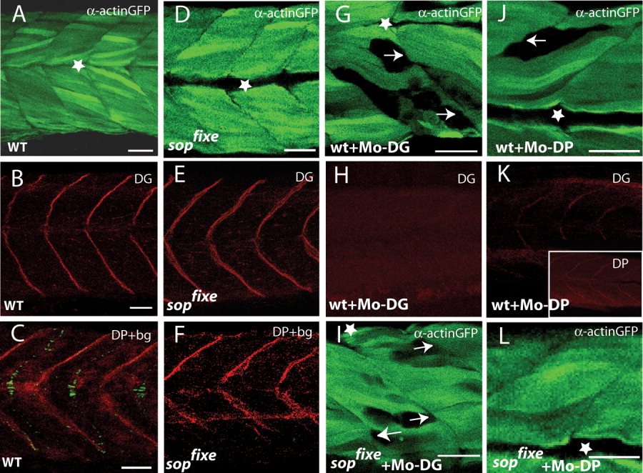
Fig. 5 sopfixe suppresses the muscle defects in embryos lacking Dystrophin. A-C: Uninjected wild-type embryos showing expression of the α-actin:GFP transgene (A) or expression of dystroglycan (DG, B) or the combination of Dystrophin (DP) immunofluorescence and α-bungarotoxin staining (C). D-F: sopfixe mutants showing expression of the α-actin:GFP transgene (D) or expression of DG (E) or the combination of DP immunofluorescence and α-bungarotoxin staining (F). Lack of α-bungarotoxin in sopfixe does not affect the pattern of DG or DP expression (compare B,C with E,F). G,H: Wild-type embryos injected with the morpholino against DG (Mo-DG). G: Removal of DG causes detachment of the α-actin:gfp labeled myofibrils from the vertical myosepta (arrows). H: The injected Mo-DG completely abolishes DG expression. I: sopfixe embryo injected with Mo-DG shows a similar pattern of muscle defects as wild-type embryos, in which DG was knocked down, indicating that sopfixe does not suppress the muscle phenotype in Mo-DG morphants. J,K: Embryos in which DP was knocked down show detachment of myofibrils (J) and also a reduction in DG as well as DP staining at the vertical myosepta (K, and insert in K, respectively). L: sopfixe mutant embryos in which DP was knocked down do not show detachment of myofibrils from the vertical myoseptum. Thus, sopfixe suppresses the muscle defects in Mo-DP morphants. A,B, D,E,G-L: 72 hours postfertilization (hpf); C,F: 24 hpf embryos. Asterisk, horizontal myosepta. GFP, green fluorescent protein. Scale bars = 30 μm in A,B,D,E,G-L; 25 μm in C,F.
Image
Figure Caption
Acknowledgments
This image is the copyrighted work of the attributed author or publisher, and
ZFIN has permission only to display this image to its users.
Additional permissions should be obtained from the applicable author or publisher of the image.
Full text @ Dev. Dyn.

