
|
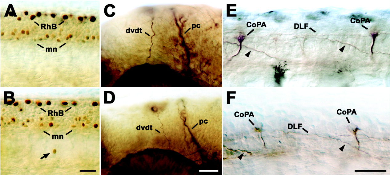
Fig. 5 Other neurons and axons in the head and spinal cord are not affected by NRP1a morpholino1. Lateral views of 24 hpf whole-mounted embryos at mid-trunk (A,B,E,F) or head (C,D) levels are shown (rostral is left). A,B: Labeling Rohon-Beard (RhB) and motor neurons (mn) with an antibody to islet-1 indicates comparable numbers of these cell types in uninjected embryos (A) and those injected with 1mM NRP1a morpholino1 (B). Whereas Rohon-Beard neurons and most motor neurons are found in their correct locations, one immunopositive cell (arrow) is displaced ventral to the spinal cord in B. C,D: Anti-tubulin labeling of the dorsoventral diencephalic tract (dvdt) and the posterior commissure (pc) reveals no significant differences between uninjected embryos (C) and those injected with 1 mM NRP1a morpholino1 (D). E,F: Labeling of commissural primary ascending interneurons in the spinal cord with the 3A10 antibody indicated normal positioning of somata (CoPA) and contralateral axons (arrowheads), which eventually join the dorsal longitudinal fascicle (DLF) in uninjected embryos (E) and those injected with 1mM NRP1a morpholino1 (F). Scale bars = 25 μm in B (applies to A,B), 25 μm in D (applies to C,D), 25 μm in F (applies to E,F).