
|
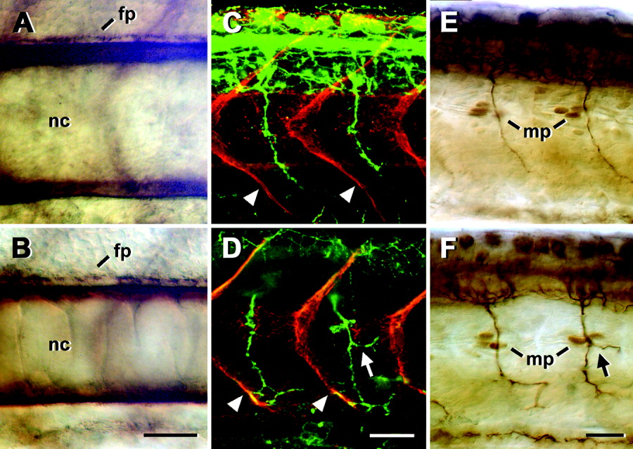
Fig. 4 Trunk structures appear normal after injection of 1 mM neuropilin-1a (NRP1a) morpholino1. Lateral views of whole-mounted 24 hours postfertilization (hpf) embryos at mid-trunk levels are shown (rostral is left). A-F: Notochord (nc) and spinal floor plate (fp) labeled with an anti-chondroitin sulfate antibody (A,B), vertical myosepta (arrowheads) labeled with an anti-tenascin-C antibody (red), and ventral motor axons labeled with an anti-HNK-1 antibody (green, C,D), as well as muscle pioneer cells (mp) at the horizontal myoseptum labeled with an antibody to engrailed, and ventral motor axons labeled with an anti-tubulin antibody (E,F) did not show systematic differences between uninjected embryos (A,C,E) and those injected with 1 mM NRP1a morpholino1 (B,D,F). Arrows in D and F indicate aberrant branches of ventral motor nerves. Scale bars = 25 μm in B (applies to A,B), 25 μm in D (applies to C,D), 25 μm in F (applies to E,F).