
|
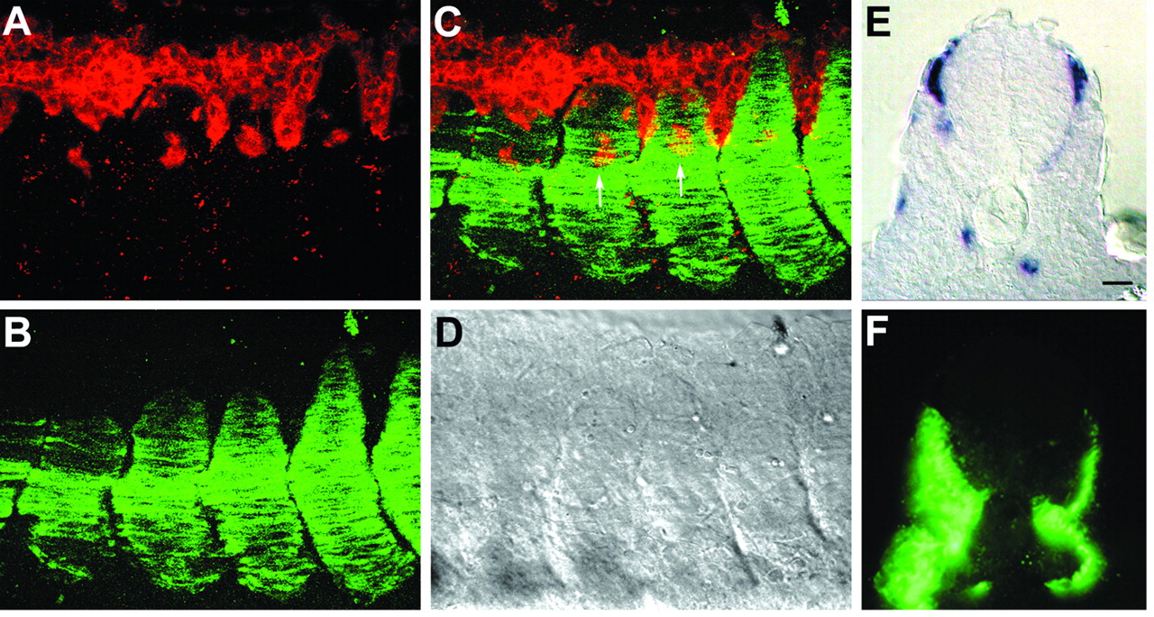
Fig. 6 Fast muscle is not essential for neural crest migration on the medial pathway. (A-D) Whole-mount staining of shh mRNA-injected embryo at 21 hpf. (A) crestin RNA expression (red) in neural crest cells. (B) F59 protein localization (green) in slow muscle. (C) Merged image. (D) DIC image. In the absence of fast muscle, the somites do not adhere to one another, creating a somite-free space in the intersomitic cleft. Neural crest migrates in this region, presumably because neural crest cells are motile in the absence of somites. However, neural crest also migrates in normal streams medial to the middle of somites (arrows in C). (E,F) Cross-section of shh mRNA-injected embryo. The somite on the left side of this embryo has no fast muscle and excess slow muscle (all the muscle is slow, as indicated by F59 staining in F); however, neural crest cells (crestin riboprobe, purple in E) have migrated to the same extent as on the right side, where fast muscle is present and slow muscle has migrated to the myotome periphery, as in wild type. Scale bar: 20 µm.