
|
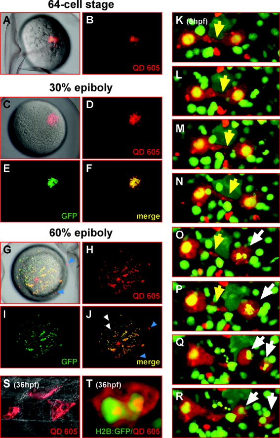
Fig. 3 Lineage tracing of zebrafish embryonic cells using QD605. A: Two blastomeres of a zebrafish embryo at the 64-cell stage (overlay of transmitted light image with QD605 fluorescence) have been coinjected with QD605 and mRNA encoding green fluorescent protein (GFP), an established lineage tracer in zebrafish embryos. B: The restriction to two individual blastomeres can be observed immediately after the injection by the red fluorescence of QD605 using a fluorescence stereomicroscope. C-E: At 30% epiboly (C, overlay of transmitted light image with QD605 fluorescence), green fluorescence emitted by the meanwhile translated GFP can be observed (E) in addition to the red QD605-derived fluorescence (D). Both fluorescent signals are localized to a single continuous cell cluster of approximately 20 cells close to the apical pole of the embryos. F: The overlay of both fluorescent channels shows that both signals overlap completely. This finding indicates that streptavidin-conjugated QD605 do not spread to neighboring cells through gap junctions but are being passed on to descendants of the initially injected blastomeres through rounds of cell divisions. G: At 60% epiboly, the single cell cluster of fluorescent cells has dispersed, likely due to extensive cell movements of gastrulating cells. H,I: Red QD605-derived fluorescence (H) and green GFP-emitted fluorescence (I) can still be detected in numerous cells. J: The overlap of both fluorescent channels shows that most fluorescent cells co-emit GFP and QD605 fluorescence, indicating that streptavidin-conjugated QD605 can be used to mark descendants of individually injected cells. Some cells appear to display green GFP fluorescence only (J, white arrowheads). The lack of QD605 in this subfraction of cells may be caused by QD605 being propagated to only one daughter cell during a round of cell division, due to their tendency to form aggregates. G,J: A few red fluorescent signals emitted from QD605 that do not co-emit GFP fluorescence appear to be derived from quantum dots that are not localized within the embryo, perhaps being deposited between the embryo and the chorion during the injection procedure (compare blue arrowheads in J and G). These results show that streptavidin-conjugated QD605 can be used as lineage tracers in zebrafish embryos to label the vast majority of descendants from an injected individual precursor cell. K-R: To directly follow individual cell dynamics, zebrafish embryos were injected with mRNA (150 ng/μl) encoding a histone2B-EGFP fusion protein at the single-cell stage followed by QD605 injection of an individual blastomere at the 64-cell stage. H: As such labeled cells disperse during gastrulation individually QD605-labeled cells could be followed by time-lapse recording using laser scanning confocal microscopy. Starting at 8 hours postfertilization (hpf) stacks of images spaced by 5 μm, respectively, were recorded every 5 min covering an optical section of 30 μm in total. A sequence from a several-hours recording is displayed with images from a 10-μm fraction of the recorded stacks being projected into single frames by maximum intensity projection. Two cells making and releasing contact (K-P, yellow arrowheads) while undergoing morphological changes can be observed due to the QD605-derived fluorescence of their cytoplasm. Subsequently, the chromatin of one of the labeled cells condenses (O,P, white arrows), and two daughter cells (Q,R, white arrows) arise from a cell division, each carrying the QD605 label. T: To record more elaborate cell morphology, a skin cell from a similarly injected embryo was imaged at 36 hpf. S: In addition, individual notochord cells were injected at 36 hpf with QD605. Pictures from a recorded stack of images (60 μm at 1 μm distance) were projected by maximum intensity projection into a single plane. Due to the bright fluorescent labeling, the polygonal shape of the notochord cell can be appreciated with the nucleus and vacuoles being spared by the fluorescent quantum dots. nc, notochord; sm, somitic muscle.