
|
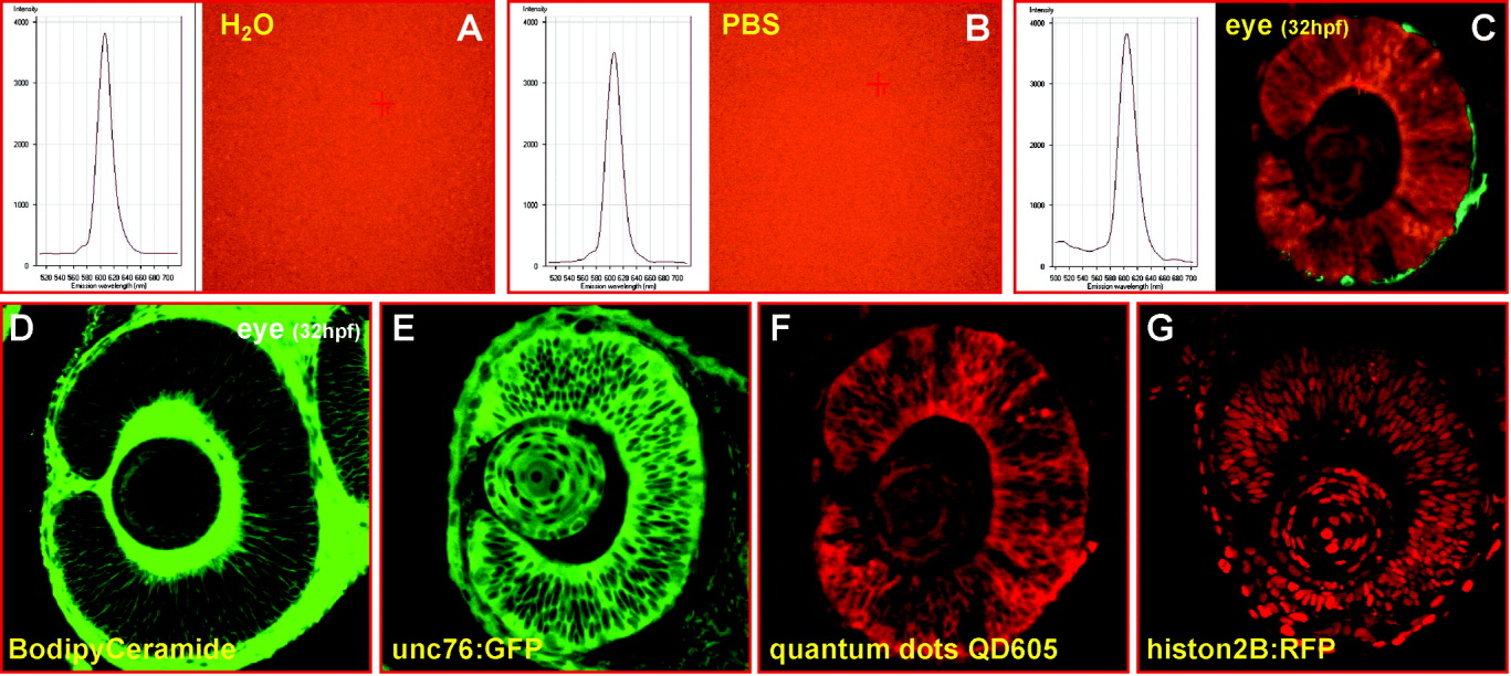
Fig. 2 Emission and localization properties of QD605. A-C: Comparison of the emission spectra of QD605 measured in water (A), phosphate buffered saline (PBS, B), and within retinal cells of zebrafish embryos (C) indicates that the QD605 emission spectra remain unaltered in vitro and in vivo irrespective of their environment. Spectroscopy was performed using laser scanning confocal microscopy combined with liquid crystal tunable filters (LSM510 Meta system, Zeiss). The cross hair demarcates the spot from which the spectra were obtained. Relocating the cross hair to different points of fluorescence did not affect the emission spectra significantly. D-G: Comparison of differently localized cellular fluorescent labels within lens fiber and retinal cells in developing zebrafish eyes using laser scanning confocal microscopy; whereas the membrane-localized dye Bodipy Ceramide (D) and the nuclear-localized histon2B-mRFP fusion protein (G) result in a different vital cellular label, the signal obtained from streptavidin-conjugated QD605 (F) appeared to resemble very closely the labeling of the unc76:GFP fusion protein, which labels the cellular cytoplasm but is excluded from the nucleus (E). hpf, hours postfertilization; RFP, red fluorescent protein.