
Image
|
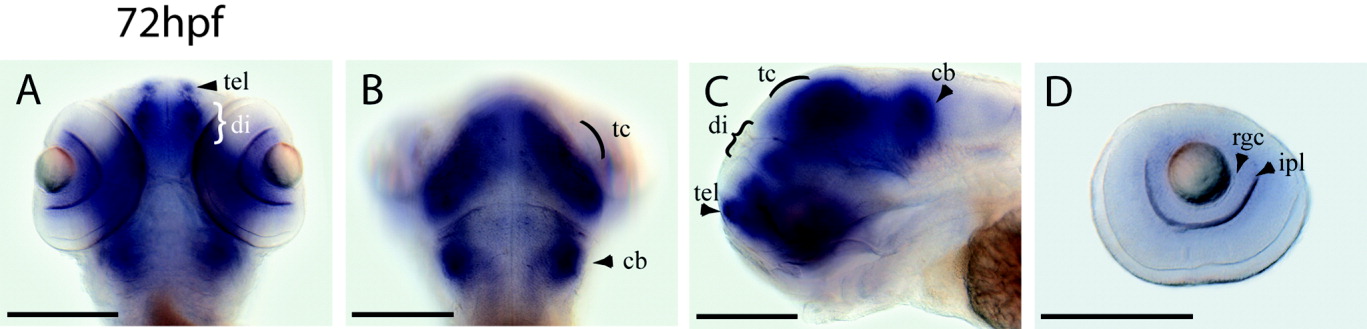
Figure Caption
Fig. 4 Whole-mount in situ hybridization analysis of foxP2 RNA expression during zebrafish development, stage 72 hpf. A,B: Dorsal views at 72 hpf in two different focal planes. C: Lateral view at 72 hpf. D: Expression in the retina at 72 hpf. Lateral views: anterior to the left; dorsal views: anterior to the top. Scale bars are 50 μm, except in D, where scale bar is 25 μm. cb, cerebellum; di, diencephalon; ipl, inner plexiform layer; rgc, retinal ganglion cells; tc, tectum; tel, telencephalon.
Figure Data
Acknowledgments
This image is the copyrighted work of the attributed author or publisher, and
ZFIN has permission only to display this image to its users.
Additional permissions should be obtained from the applicable author or publisher of the image.
Full text @ Dev. Dyn.