
|
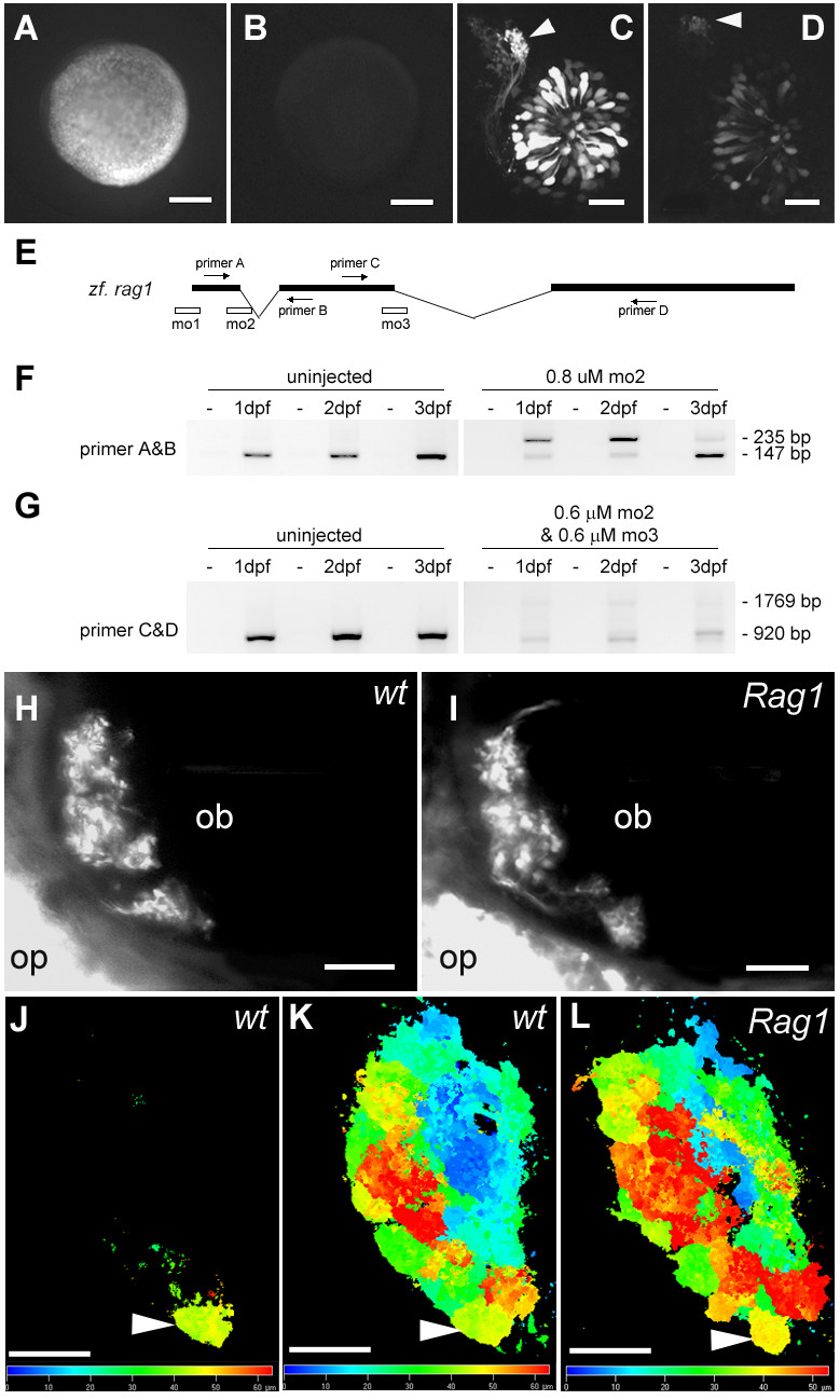
Fig. 5 The effect of RAG1 depletion on the olfactory projection. (A) An embryo injected with mRNA encoding the 5' end of Rag1 fused to EGFP. (B) An embryo co-injected with the Rag1-EGFP fusion mRNA and mo1. (C) Olfactory neurons labelled with GFP under the Rag1 promoter, with brightly labeled axons projecting to a single target (arrowhead), at 3 dpf. (D) In a transgenic embryo injected with mo1, axons still project to the same target (arrowhead), but the intensity of GFP fluorescence is reduced. (E) A schematic diagram of the Rag1 gene, showing the location of morpholinos and primers that were used to analyse morpholino-injected fish. (F) RT-PCR on control or mo2 injected embryos. Abnormal splicing occurs in the morpholino-injected fish, leading to a premature stop codon, as indicated by sequencing of the upper band. (G) RT-PCR after injection of a mixture of Rag1 mo2 and mo3, showing loss of the normal transcript. (H, I) A subset of Di8ANEPPQ-labeled olfactory sensory neurons in 7-day old wild type and Rag1 mutant fish. Axons innervate all target structures detectable in this optical plane in the mutant. (J-L) SV2-labelled 4 day-old Rag1:GFP transgenic (J, K) and Rag1 mutant (L) forebrains, shown in dorsal view. The images are colour-coded according to depth. The glomerulus innervated by the strong GFP-positive neurons (J) is indicated by the arrowhead. Bar = 100 µm (A, B); 20 µm (C, D, H-K). The colour bars in J and K indicate depth. Embryos in panels H-L are shown in dorsal view, with anterior to the left.