
Image
|
Figure Caption
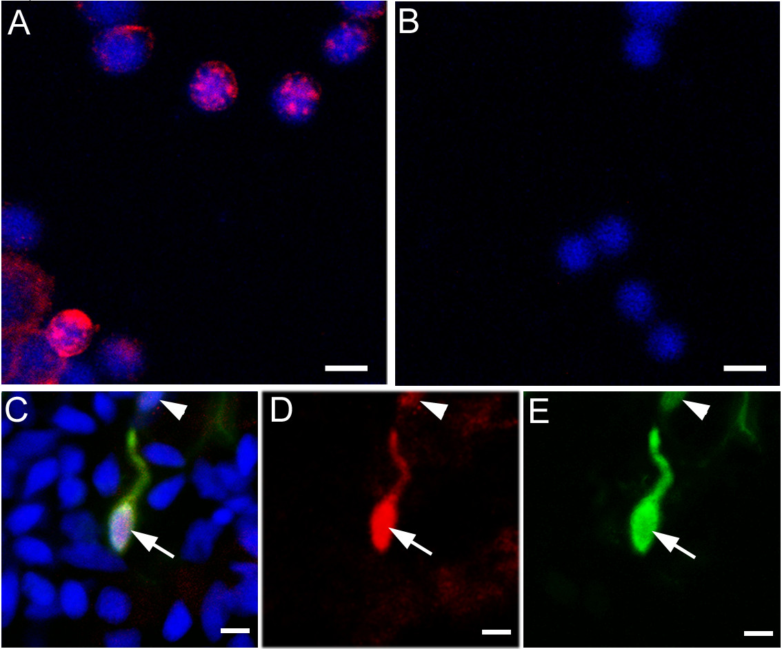
Fig. 1 Immunofluorescent labelling of RAG1. (A) Isolated zebrafish thymocytes, labelled with the antibody to RAG1 (in red) and DAPI (blue). (B) After pre-absorption with the peptide used for immunization, no labelling was detected. (C-E) Double-label of olfactory epithelial cells isolated from a Rag1:GFP transgenic fish. RAG1 protein (C, D; red) is present in the GFP-positive (C, E; green) neurons (arrow and arrowhead). Neurons with high (arrow) or low (arrowhead) GFP levels contain RAG1. DAPI (C; blue) is used to stain nuclei. Bar = 5 µm.
Figure Data
Acknowledgments
This image is the copyrighted work of the attributed author or publisher, and
ZFIN has permission only to display this image to its users.
Additional permissions should be obtained from the applicable author or publisher of the image.
Full text @ BMC Neurosci.