
|
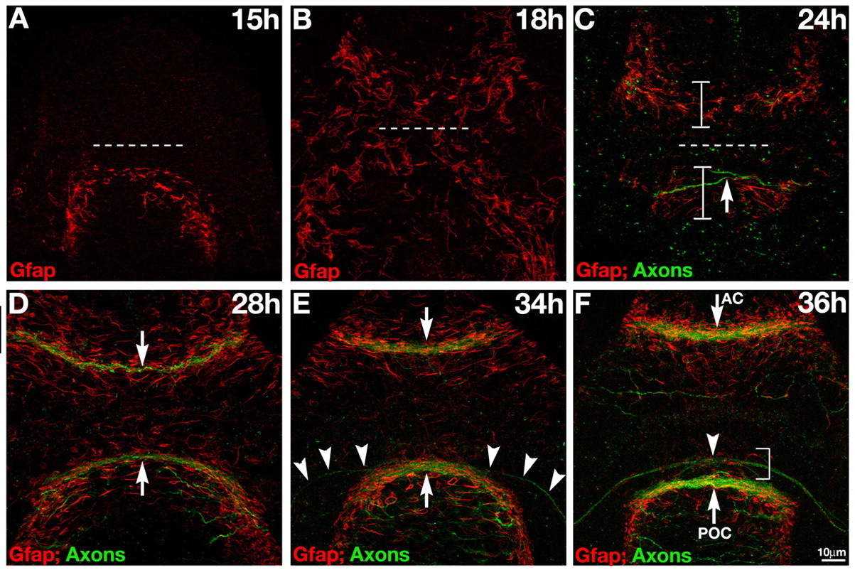
Fig. 2 Gfap+ cells span the midline prior to formation of forebrain commissures. (A-F) Double labeling for axons (anti-AT; green) and Gfap+ cells (red). (A) Gfap+ cells span the midline posterior to the optic recess (dashed line) at the presumptive POC at 15 hpf. (B) By 18 hpf, Gfap+ cells span the midline, anterior and posterior to the optic recess (dashed line), at the presumptive AC and POC. (C) By 24 hpf, more distinct glial bridges (brackets) have formed at the presumptive AC and the forming POC (arrow). (D) At 28 hpf both commissures (arrows) have formed in association with the glial bridges. (E) The first RGC axons (arrowheads) grow towards the midline along Gfap+ cells at 34 hpf. (F) The optic chiasm (arrowhead) forms in association with a distinct cluster of Gfap+ fibers just anterior to the POC (bracket). At 15 hpf and 18 hpf, no axons are present in the forebrain (these data) (Wilson et al., 1990), so the green channel was omitted to reduce background. All panels show frontal views of the forebrain. Scale bar: 10 μm.