
|
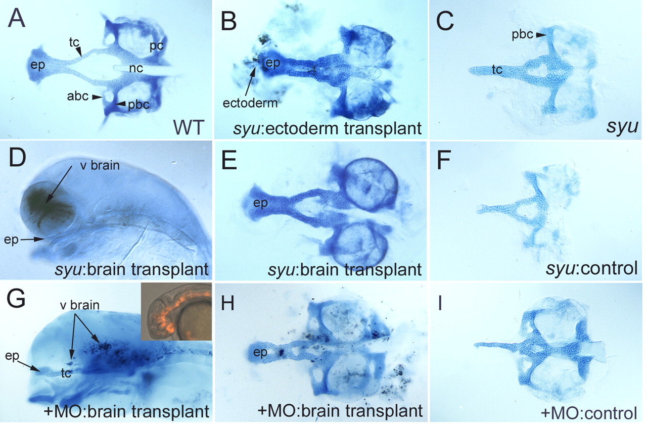
Fig. 8 Shh is required in both ventral neural tube and oral ectoderm for ANC formation. (A-C,E,F,H,I) Flat-mounted Alcian-Blue stained cartilage at 5 dpf; anterior is to the left. (D,G) Lateral views of transplants shown in E and H, prior to cartilage dissection. Transplanted donor cells (brown) lie in the ventral brain. (A) Wild type. (B) Partial rescue of the ANC by wild-type ectoderm transplanted into a syu mutant host. Transplanted cells lie close to the ethmoid plate in this example. (C) syu control sibling of the embryo shown in B. (D) Grafted wild-type cells in the ventral forebrain of a syu mutant. (E) Partial rescue of the ANC by the transplant shown in D. Grafted cells were lost during dissection. (F) syu control sibling of embryo shown in E. (G) An shh-MO/twhh-MO-injected embryo. Inset shows transplanted cells at 28 hpf. (H) Partial rescue of ANC in the embryo shown in G. (I) shh-MO/twhh-MO-injected control sibling of embryo in H. abc, anterior basicapsular commissure; ep, ethmoid plate; nc, notochord; pbc, posterior basicapsular commissure; pc, parachordal cartilage; tc, trabeculae.