
|
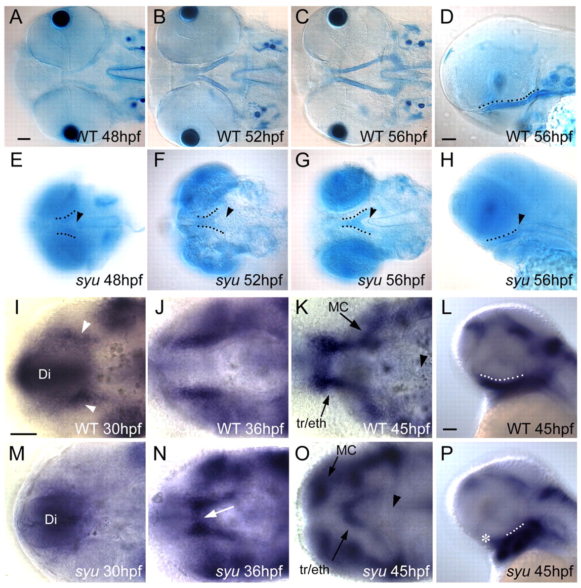
Fig. 2 Early chondrogenesis in the ANC. (A-H) Alcian Blue-stained embryos. (I-P) In situ hybridization for sox9a mRNA. Ventral views in wild type (A-C) and in syu mutants (E-G) between 48-56 hpf are shown. (A) Paired trabeculae consist of single rows of chondrocytes. (B,C) These elongate (B) and fuse posteriorly (C) to the parachordals. (D) Lateral view, 56 hpf, showing the ventral position of trabeculae (dotted line). (E) In syu mutants, the first ANC cartilages (dotted lines) are shorter and closer to the notochord (arrowhead). (F) Trabeculae fuse at the midline and (G) this fusion persists. (H) A lateral view of a syu mutant at 56 hpf. (I-K) Ventral views of sox9a expression in wild type between 30-45 hpf. (M-O) sox9a expression in syu mutants. (I) Early expression in bilateral cell clusters (arrowheads) adjacent to the diencephalon (Di). (J,K) These elongate posteriorly (J), and fuse in the midline anteriorly (K). (L) Lateral view of wild type at 45 hpf. (M) In syu mutants, sox9a expression is delayed. (N,O) sox9a+ clusters elongate (N) and fuse anteriorly (O, arrow). (P) Lateral view of sox9a expression in syu mutants showing fused trabeculae (dotted lines). Asterisk indicates anterior extent of expression. eth, ethmoid plate; MC, Meckel's cartilage; tr, trabeculae. Scale bar: 50 μm.