
|
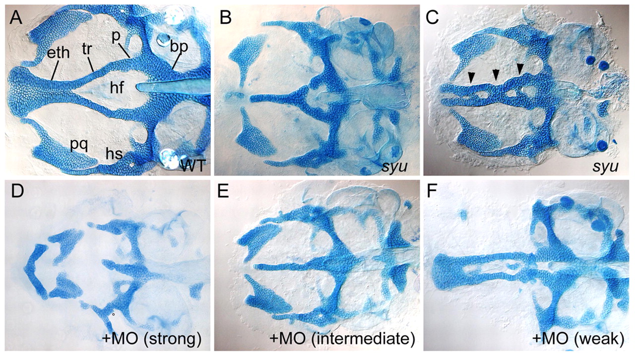
Fig. 1 Neurocranial cartilage patterns in wild-type and Hh-deficient larvae. Alcian stained cartilages were dissected and flat mounted; dorsal views are shown, anterior to the left. (A) The wild-type ANC at 4.5 dpf includes paired trabeculae (tr) and an ethmoid plate (eth). Mandibular (pq) and hyoid (hs) cartilages remain attached. (B,C) Neurocranial defects in syu mutants. Trabecular cartilages either fuse completely (B) or partially (C, arrowheads) at the midline. (D-F) Concentration-dependent effects of co-injected shh-MO and twhh-MO in wild type: (D) 3.0 ng shh-MO + 0.82 ng twhh-MO; (E) 2.0 ng shh-MO + 0.55 ng twhh-MO; (F) 1.0 ng shh-MO + 0.27 ng twhh-MO. bp, basal plate; eth, ethmoid plate; hf, hypophysial fenestra; hs, hyosymplectic; pq, palatoquadrate; p, polar cartilage; tr, trabeculae.