
Image
|
Figure Caption
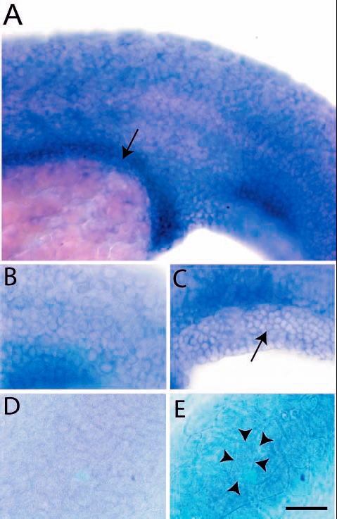
Fig. 5 Expression analysis of lgl2 by in situ hybridisation. Epidermal cells and presumptive gut (arrow) of a 24-hour-old larva exhibit lgl2 expression (A). The basal cells of epidermis (B) including fin fold epidermis (C, arrow) express lgl2. Basal epidermis (D) and periderm (E) of control embryo hybridised with sense probe. The peridermal cells (cell borders marked by arrowheads in E) are bigger than the basal epidermal cells (B,C). Scale bar: 50 µm in A; 33 µm in B-E.
Figure Data
Acknowledgments
This image is the copyrighted work of the attributed author or publisher, and
ZFIN has permission only to display this image to its users.
Additional permissions should be obtained from the applicable author or publisher of the image.
Full text @ Development