
|
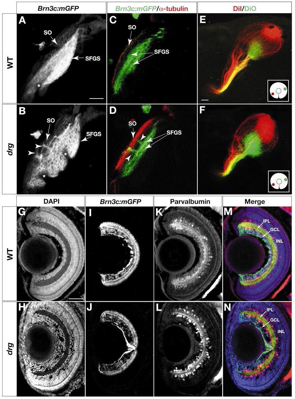
Fig. 6 Laminar specificity is perturbed in dragnet (drg) mutants. (A-D) Analysis of retinorecipient layers in 6 dpf wild type (A,C) and drg (B,D). (A,B) Z projections of confocal image stacks, labeled with Brn3c:mGFP. Larvae are mounted at a slightly oblique angle to better visualize the gap between SO and SFGS. (C,D) Optical sections of Brn3c:mGFP tecta, stained with whole-mount anti-acetylated tubulin (red) and anti-GFP (green). Arrowheads indicate ectoptic axon fascicles traveling between SO and SFGS in drg mutants. (E,F) Analysis of the retinotopic map in 6 dpf wild type (E) and drg (F). DiI (red) and DiO (green) were pressure injected into ventronasal and dorsotemporal retina, respectively (see inserts for illustration of injection sites). Axon targeting in drg (F) is comparable with wild type (E), suggesting that positional information along the retinotopic axes is intact. (G-N) Analysis of the inner plexiform layer (IPL) in sections of 6 dpf retina (see M,N for labels of retinal layers). (G,H) DAPI labeling. (I,J) Anti-GFP labeling to visualize the four sublaminae to which Brn3c:mGFP RGC dendrites project. (K,L) Anti-parvalbumin labeling, to highlight a subpopulation of amacrine cells that project to three sublaminae in the IPL. (M,N) Triple-labeling (merged image) showing DAPI (blue), anti-GFP (green) and anti-parvalbumin (red). Formation of IPL sublaminae is not affected in drg. An apparently unrelated phenotype of drg can be seen using the DAPI stain: an abnormal aggregation of cells in front of the drg lens (H). Asterisks indicate melanophores in the skin. Scale bars: 20 µm in A,G.