
|
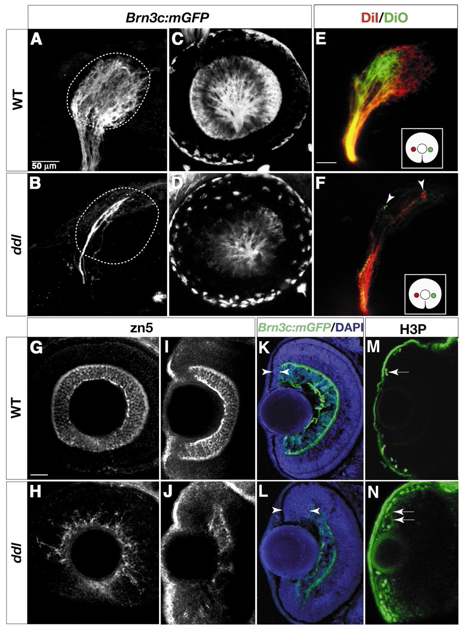
Fig. 3 daredevil (ddl) mutants have fewer RGCs. Analysis of cell-type markers in wild type (A,C,E,G,I,K,M) and ddl (B,D,F,H,J,L,N). (A,B) Lateral views of 78 hpf tecta, labeled with whole-mount anti-GFP. Broken lines outline boundaries of the tectal neuropil. The wild-type tectum (A) is covered by axons. Few axons can be detected in the ddl tectum (B). (C,D) Confocal images of retinas in live 60 hpf embryos. The number of GFP-positive RGCs is greatly reduced in ddl (D). (E,F) Analysis of the retinotopic map in 72 hpf wild type (E) and ddl (F). DiI (red) and DiO (green) were pressure injected into nasal and temporal retina, respectively (see inserts for illustration of retinal injection sites). The gross topography of axon targeting in ddl mutants is not affected, with nasal axons still projecting to the posterior tectum and temporal axons to the anterior tectum (F). (G-J) Whole-mount Zn5 staining of 72 hpf retinas. (G,H) Lateral views. (I,J) Dorsal views. The number of zn5-positive RGCs is greatly reduced in ddl. (K,L) Sections of 78 hpf retinae, labeled with anti-GFP (green) and DAPI (blue). The ciliary margin (between arrowheads) in the ddl retina is wider than in wild type. (M,N) Dorsal views of 72 hpf whole-mount retinae, labeled with anti-phosphohistone H3 (H3P). The number of dividing cells (arrows) is greatly increased at the ciliary margin in ddl. Scale bars: 50 µm in A and E (for tectum panels); 20 µm in G (for retina panels).